Figures & data
Table 1. Association between PD-L1 expression and clinicopathological features of nasopharyngeal carcinoma patients (N = 209)
Figure 1. Evaluation of CD3, PD-L1 protein and their associations in nasopharyngeal carcinoma patients. Tumor and immune cell PD-L1 protein (A) and intratumoral and peritumoral CD3+ lymphocytes (B). Correlation between intratumoral (C) or peritumoral CD3+ lymphocytes and TCs-PD-L1 (D), and correlation between intratumoral (E) or peritumoral CD3+ lymphocytes (F) and ICs- PD-L1 expression.
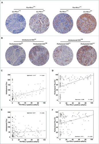
Table 2. Cox proportional regression analysis for the prediction of nasopharyngeal patient OS and DFS
Figure 2. Survival curves of different distributions of CD3+ TILs. Kaplan–Meier plots of OS (A) and DFS (B) according to the percentage of TILs. OS (C and E) and DFS (D and F) based on intratumoral and peritumoral CD3+ lymphocytes.
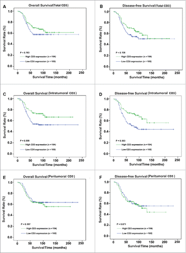
Figure 3. Survival curves of different distributions of PD-L1+ cells. The present study analyzed OS (A and C) and DFS (B and D) based on TCs-PD-L1 and ICs-PD-L1.
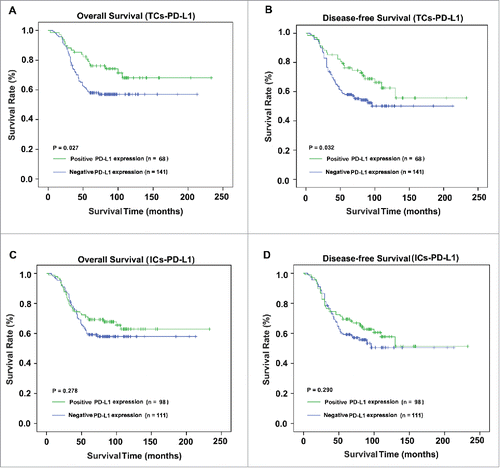
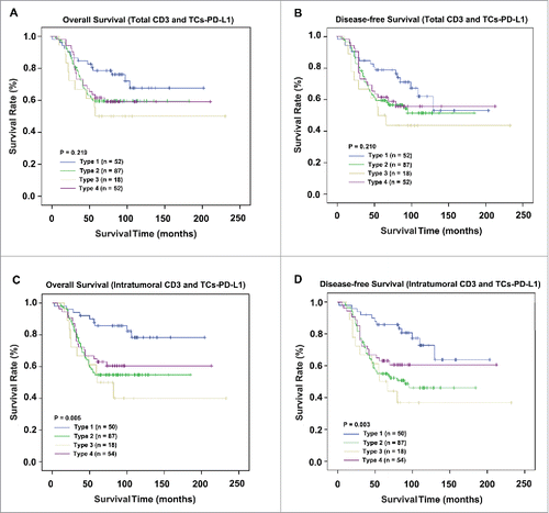
Figure 4. Comparison of survival according to staging system in 209 NPC patients. OS and DFS analyses according to the stage classification based on CD3+ lymphocytes and TCs-PD-L1 (A and B). Survival analysis based on intratumoral CD3+ lymphocytes and TCs-PD-L1 (C and D).