Figures & data
Figure 1. (A) XRT treatment, lymphocyte assessment and lymph node harvest scheme for 4 Gy x 10 fractions(abbreviated) and 2 Gy x 30 fractions(prolonged) groups. (B) Visual representation of treatment scheme and animal strains used in this study. (C) Imaging display from SARRP showing that radiation treatment is extremely precise and that peripheral sites are not exposed to XRT. (D) Focal alopecia is induced in treated animals, highlighting the focused treatment and successful XRT exposure.
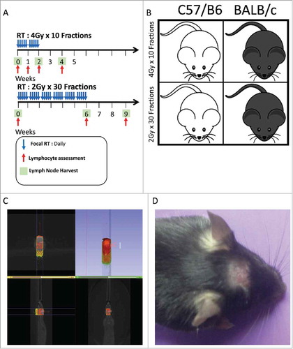
Figure 2. (A) Lymphocyte assessment for the C57 BL/6 strain treated with the 4 Gy x 10 fractions, showing non-specific lymphodepletion in the periphery as response to treatment. (B) Lymphocyte assessment for the Balb/c strain treated with the 4 Gy x 10 fractions, showing non-specific lymphodepletion in the periphery as response to treatment. All experiments were repeated in triplicate. Error bars were calculated using one-way repeated measures analysis of variance. Middle bars represent mean, with upper and lower error bars.
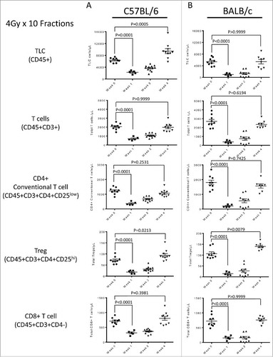
Figure 3. (A) Lymphocyte assessment for the C57 BL/6 strain treated with the 2 Gy x 30 fractions, showing non-specific lymphodepletion in the periphery as response to treatment. (B) Lymphocyte assessment for the Balb/c strain treated with the 2 Gy x 30 fractions, showing non-specific lymphodepletion in the periphery as response to treatment. All experiments were repeated in triplicate. Error bars were calculated using one-way repeated measures analysis of variance. Middle bars represent mean, with upper and lower error bars.
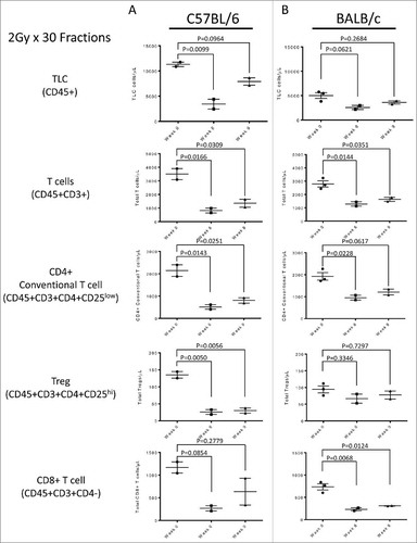
Figure 4. (A) Baseline lymph node assessment for the C57 BL/6 strain treated with the 4 Gy x 10 fractions at 40X and 160X magnification, showing typical LN at baseline. (B) End of treatment lymph node assessment for the C57 BL/6 strain treated with the 4 Gy x 10 fractions. Lymphoid architecture is distorted by a paracortical expansion with a mixed lymphohistiocytic composition (40X, *) and there are prominent aggregates of hilar histiocytes (160X). (C) Recovery lymph node for the C57 BL/6 strain treated with the 4 Gy x 10 fractions. Vaguely nodular, histiocyte-rich paracortical expansions remain prominent (40X, *) with some histiocytes and fibrosis adjacent to organized lymphoid follicles (160X).
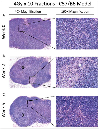
Figure 5. (A) Baseline lymph node assessment for the C57 BL/6 strain treated with the 2 Gy x 30 fractions at 40X and 160X magnification, showing typical architecture at baseline. Cortical areas and paracortical areas are lymphocyte-rich. (B) End of treatment lymph node assessment for the C57 BL/6 strain treated with the 2 Gy x 30 fractions at 40X and 160X magnification. Paracortical, predominantly histiocytic expansions are prominent (*) and lymphocytes are relatively restricted to follicles (**). (C) Recovery lymph node assessment for the C57 BL/6 strain treated with the 2 Gy x 30 fractions at 40X and 160X. Paracortical expansions persist with fibrosis (60X, *) and hilar epitheloid histiocytes and scattered sea-blue histiocytes are prominent. Tingible body macrophages, and pigment-laden macrophages indicating a dermatopathic component to the lymph node changes, were also present (not shown).
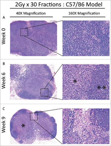
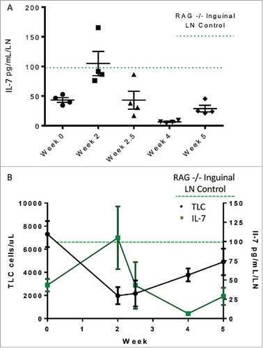
Figure 6. (A) IL-7 levels in distal lymph nodes measured over the course of treatment in C57 Bl/6 animals treated with 4 Gy x 10 fractions. (B) IL-7 levels are inversely correlated with peripheral lymphocyte counts.