Figures & data
Figure 1. Vitiligo confers an intermediate protection against melanoma development compared to NOS2 deficiency.
(A) Time courses of vitiligo onset from Ret (n = 40) and RetNos2KO (n = 40) mice. (B-C) 6-months follow-up of melanoma development from Ret mice without vitiligo (RetNV, n = 24), Ret mice with vitiligo (RetV, n = 16) and RetNos2KO (n = 40) mice. Time courses of primary tumor (B) and cutaneous metastasis (C) onset. Mice were examined every two weeks. *P < 0.05 and ****P < 0.0001 (Wilcoxon test).
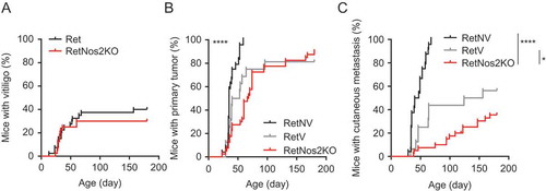
Figure 2. Vitiligo-associated decrease of PMN-MDSCs and γδ T cell infiltration within primary tumors and spleen.
(A) Protein levels of indicated cytokines in aqueous humors from RetNV (n = 14) and RetV (n = 11) mice, determined by multiplex ELISA. (B) IL-1β and IL-6 quantifications in sera from RetNV (n = 8) and RetV (n = 8) mice determined by ELISA. (C-F) Dendritic cells (CD11c+), macrophages (CD11b+CD11c−Ly6C−/lowLy6G−), monocytic MDSCs (M-MDSCs: CD11b+CD11c−Ly6ChighLy6G−), PMN-MDSCs (CD11b+CD11c−Ly6ClowLy6G+), CD4+ T cells (CD3+, CD4+), CD8+ T cells (CD3+, CD8+), γδ T cells (CD3+, δ TCR+), NK cells (CD3−, NK1.1+), B cells (CD3−, CD19+) were analyzed by flow cytometry. Parts of whole graphs describing immune cells found in primary tumors (C). Percentages of CD8+ T cells, CD4+ T cells, NK cells and B lymphocytes (D) PMN-MDSCs and γδ T cells among CD45+ cells within primary tumors (E) from RetNV (n = 18) and RetV (n = 19) or within spleen (F) from RetNV (n = 7) and RetV (n = 16). Each point represents individual mouse. Bars are mean ± SEM. *P < 0.05, **P < 0.01 ***P < 0.001 (Mann-Whitney test).
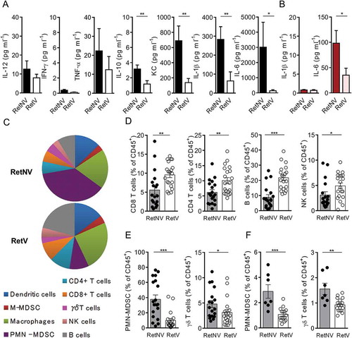
Figure 3. Vitiligo-associated decrease of the proportion of NOS2 positive γδ T cells in primary melanoma and TdLNs.
(A) Representative microscopy images showing γδ T cells positive for NOS2 from cells derived from TdLNs of RetNV or RetV mice and stained with antibodies to TCR γδ (red), NOS2 (green) and counterstained with DAPI (blue). Arrows indicate NOS2 expressing γδ T cells. Bars 10 µM. 40 X objective. (B, C) Quantification of NOS2 positive γδ T cells in TdLN (B) from RetNV (n = 7) and RetV (n = 6) and in primary melanoma (C) from RetNV and RetV (n = 4 each). It was performed from 500 to 1500 γδ T cells. Each point represents individual mouse. Bars are mean ± SEM. *P < 0.05, **P < 0.01 (Mann-Whitney test).
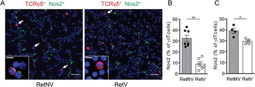
Figure 4. In vivo neutralization of IL-1β and IL-6 slows down metastasis formation and reduced PMN-MDSCs and γδ T cell infiltration within primary tumors and spleen.
(A, B) 3-month follow-up of melanoma development from Ret mice untreated (n = 12) or treated either with antibodies neutralizing IL-1β and IL-6 (n = 12) or with IL-1 receptor antagonist (anakinra) (n = 14). Time courses of primary tumor (A), cutaneous metastasis (B) and vitiligo (C) onsets. (D) Protein levels of IL-1β and IL-6 in primary tumor extracts, from Ret (n = 4) and Ret treated (n = 4) mice, determined by multiplex ELISA. (E-H) Analyses of immune cell proportions within primary tumors (E) and spleen (G) represented as parts of whole. Percentages of PMN-MDSCs and γδ T cells among CD45+ cells within primary tumors (F) or spleen (H) from Ret (n = 9 and 8) and Ret treated (n = 7 and 11 respectively) mice. Each point represents individual mouse. Bars are mean ± SEM. *P < 0.05, **P < 0.01. Wilcoxon test (A, B), Mann-Whitney test (D, F, H).

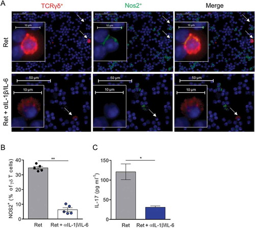
Figure 5. In vivo neutralization of IL-1β and IL-6 suppresses NOS2 expression in γδ T cells.
(A-C) 3 month old Ret mice were treated or not during 2 weeks with antibodies neutralizing IL-1β and IL-6. (A, B) NOS2 expression was analyzed by microscopy in TdLNs from 5 mice in each group. Representative images showing γδ T cells positive for NOS2 derived either from untreated or treated Ret mice and stained with antibodies to TCR γδ (red), NOS2 (green) and counterstained with DAPI (blue). Bars 10 µM. 40 X objective. Arrows indicate γδ T cells. (B) NOS2 positive γδ T cells were quantified from 500 to 1500 γδ T cells. (C) Protein levels of IL-17 in primary tumor extracts, from untreated (n = 5) and treated (n = 5) Ret mice, were determined by ELISA. Each point represents individual mouse. Bars are mean ± SEM. *P < 0.05, **P < 0.01. (Mann-Whitney test).