6,227
Views
11
CrossRef citations to date
0
Altmetric
Original Research

A cell-engineered system to assess tumor cell sensitivity to CD8+ T cell-mediated cytotoxicity

, , , , , , , , & show all
Pages 1-10 | Received 03 Oct 2018, Accepted 18 Mar 2019, Published online: 26 Apr 2019

Figures & data

Figure 1. Optimizing CD8+ T cells for tumor cell cytotoxicity assays.
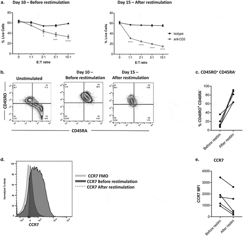
a. Percentages of live P815 target cells in the presence of soluble anti-CD3 antibody or isotype control antibody in 4-h co-cultures with CD8+ T cells before (left, day 10 after initial activation with anti-CD3/CD28 dynabeads) and after (right, day 15) a second round of stimulation with anti-CD3/CD28 dynabeads (performed on day 10). b. CD45RO and CD45RA expression by flow cytometry on naïve, unstimulated CD8 + T cells, CD8+ T cells on day 10 before and day 15 after restimulation c. Percentages of CD45RO+CD45RA CD8+T cells across multiple donors, before and after restimulation by flow cytometry. d. Representative histogram showing CCR7 expression by flow cytometry in CD8 + T cells before and after restimulation. e. MFI of CCR7 expression in CD8+ T cells across multiple donors, before and after restimulation. Data is representative of two donors from three independent experiments (mean ± SEM). E: T ratio = effector: target ratio; MFI = Mean Fluorescence Intensity; FMO = fluorescence minus one control. *P < 0.05, and ***P < 0.0005 and ****P < 0.0001, significant difference of % live cells between isotype and anti-CD3 at each E: T ratio, assessed by two-way ANOVA and Sidak‘s multiple comparison test.
Figure 1. Optimizing CD8+ T cells for tumor cell cytotoxicity assays.

Figure 2. Generation of anti-CD3 expressing tumor cell lines.
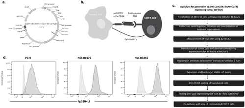
a. Schematic of anti-CD3-scFV-CD14 expression construct design. b. Schematic of anti-CD3-scFV-CD14 expressing engineered tumor cells activating CD8+ T cells via the T cell receptor (TCR), leading to T cell-mediated cytotoxicity c. Workflow of experimental steps to generate stable anti-CD3-scFV-CD14 expressing tumor cell lines for use in co-culture assays with activated CD8+ T cells. d. Representative histograms showing anti-CD3 expression on parental (light grey histograms) and transduced (dark grey histograms) PC-9, NCI-H1975 and NCI-H3255 EGFRm NSCLC cell lines, as measured by flow cytometry after surface staining with anti-IgG. Data are representative of three independent experiments. MOI = multiplicity of infection; EGFRm = epidermal growth factor receptor mutant; NSCLC = non-small cell lung carcinoma.
Figure 2. Generation of anti-CD3 expressing tumor cell lines.

Figure 3. Co-culture of CD8+ T cells with tumor cells expressing anti-CD3 results in target cell T cell-mediated cytotoxicity.
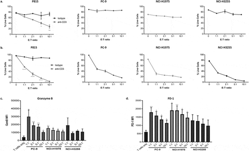
a. Percentages of live P815 cells (in the presence of soluble anti-CD3 antibody or isotype control antibody) and PC-9, NCI-H1975 and NCI-H3255 anti-CD3 engineered EGFRm cell lines in co-cultures with restimulated CD8+ T cells for 4 h. b. Percentages of live P815 cells and anti-CD3 expressing EGFRm cell lines in co-cultures with restimulated CD8+ T cells for 18 h. MFI of c. Granzyme B expression and d. PD-1 expression in CD8+ T cells alone and in co-cultures with anti-CD3 expressing PC-9, NCI-H1975 and NCI-H3255 EGFRm cell lines. Data are representative of two donors from three independent experiments (mean ± SEM). EGFRm = epidermal growth factor receptor mutant; GzmB = granzyme B; MFI = Mean Fluorescence Intensity. *P < 0.05, **P < 0.01, ***P < 0.0005 and ****P < 0.0001, significant difference of % live cells between isotype and anti-CD3 at each E: T ratio () and MFI of granzyme B and PD-1 between T cells only and at each E: T ratio (), assessed by two-way ANOVA with Sidak‘s multiple comparison test and unpaired parametric two-tailed t-test, respectively.
Figure 3. Co-culture of CD8+ T cells with tumor cells expressing anti-CD3 results in target cell T cell-mediated cytotoxicity.

Figure 4. Expression levels of anti-CD3 influence the extent of tumor cell cytotoxicity by T cells.
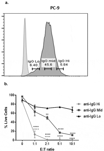
a. Representative histograms showing IgG hi, mid and lo gates on EGFRm PC-9 anti-CD3 expressing cells (dark grey histogram) compared to FMO controls (light grey histogram), using flow cytometry staining with anti-IgG. b. Percentages of live PC-9 anti-CD3 transduced cell lines when gating on IgG Hi, mid and lo tumor cell expression after 18h co-cultures with restimulated CD8+ T cells. Data are representative of two donors from three independent experiments (mean ± SEM). EGFRm = epidermal growth factor receptor mutant; FMO = fluorescence minus one control. **P < 0.01 and ****P < 0.0001, significant difference of % live cells between anti-IgG Mid vs anti-IgG lo anti-IgG Hi vs anti-IgG lo at each E: T ratio, assessed by two-way ANOVA and Sidak‘s multiple comparison test.
Figure 4. Expression levels of anti-CD3 influence the extent of tumor cell cytotoxicity by T cells.

Figure 5. Measurements of IFN-ɣ and early tumor cell apoptotic events provide additional sensitive pharmacodynamic endpoints in this assay system.
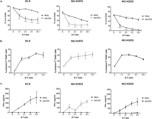
a. Percentages of live mock control cells versus anti-CD3 expressing PC-9, NCI-H1975 and NCI-H3255 EGFRm cell lines after 18h co-cultures with restimulated CD8+ T cells b. Percentages of annexin-V+ 7-AAD (apoptotic) anti-CD3 expressing PC-9, NCI-H1975 and NCI-H3255 EGFRm cell lines after 18h co-cultures with CD8+ T cells. c. Levels of IFN-ɣ production in supernatants from a. after 18h co-cultures of CD8+ T cells with mock and anti-CD3 expressing PC-9, NCI-H1975 and NCI-H3255 EGFRm tumor cell lines. Data are representative of two donors from three independent experiments (mean ± SEM). EGFRm = epidermal growth factor receptor mutant. *P < 0.05, ***P < 0.0005 and ****P < 0.0001, significant difference of % live cells between mock and anti-CD3 at each E: T ratio, assessed by two-way ANOVA and Sidak‘s multiple comparison test.
Figure 5. Measurements of IFN-ɣ and early tumor cell apoptotic events provide additional sensitive pharmacodynamic endpoints in this assay system.

Figure 6. Investigating the impact of targeted therapies on T cell-mediated cytotoxicity using the anti-CD3-expressing tumor cell co-culture system.

Percentages of live PC-9 EGFRm anti-CD3 expressing cells after 18-h co-culture with restimulated CD8+ T cells a. with PC-9 pre-treatment with 360 nM Geftinib and 160 nM Osimertinib EGFR TKIs b. in the presence of 10 µM SCH68261 and c. in the presence of 12.5 nM Dasatinib, compared to DMSO controls. Data are representative of two donors from two independent experiments (mean ± SEM), with two different donors used in each drug treatment experiment. EGFRm = epidermal growth factor receptor mutant; EGFR TKIs = epidermal growth factor receptor tyrosine kinase inhibitors; DMSO ctrl = DMSO control. **P < 0.01, and ***P < 0.0005 and ****P < 0.0001, significant difference of % live cells between drug treatments and DMSO ctrl at each E: T ratio, assessed by two-way ANOVA and Sidak‘s multiple comparison test.
Figure 6. Investigating the impact of targeted therapies on T cell-mediated cytotoxicity using the anti-CD3-expressing tumor cell co-culture system.
Supplemental material

Supplemental Material

Download MS Word (574.7 KB)