
Figures & data
Figure 1. ISG product levels predict CR after ILP
The serum concentration of (a) CXCL10, (b) CCL2, (c) PD-L2 and (d) IFN-γ in melanoma patients achieving a complete response (CR) and not achieving a complete response (Non-CR) before (pre-op.) and 1 month after (post-op.) ILP (non-paired Mann-Whitney test). The serum concentration of (e) CXCL10, (f) CCL2, (g) PD-L2 and (h) IFN-γ in melanoma patients and in healthy controls (Ctrl) (paired Wilcoxon test between pre-op. and post-op., non-paired Kruskal-Wallis test followed by Dunn’s multiple comparison test for ctrl vs. pre-op. and vs. post-op). Data are presented in box-and-whiskers plots with min. and max. (i,j) Patients were dichotomized by above or below median levels of CXCL10, CCL2, PD-L2 and IFN-γ and were grouped based on having above median levels of 0–1 ISG products, or 2–4 ISG products. The fraction of patients within each group achieving CR or not (non-CR) are shown for ISG-grouping based on (i) pre-op. samples (n = 38) and (j) post-op. samples (n = 11; Fisher’s exact test).
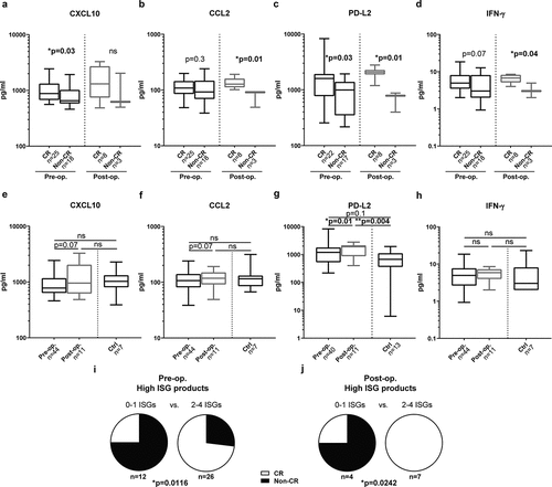
Figure 2. High levels of intratumoral ISG transcripts predict prolonged survival in advanced melanoma
Melanoma patients in the TCGA database were dichotomized by above or below median mRNA expression of the ISGs (a) CXCL10, (b) CCL2, (c) PDCD1LG2, (d) IFNG or (e) 0–1 or 2–4 of these ISGs, followed by analysis of overall survival by the log-rank test (n = 470).
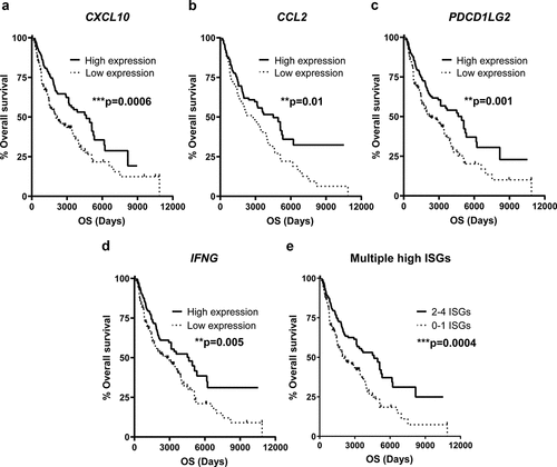
Figure 3. ILP causes induction of receptors for ISG products on PBMCs
The expression of (a–c) CXCR3 (d–f) CCR4 and (g–i) CCR5 were measured on (a, d and g) NK cells (b, e and h) CD4+ T cells and (c, f and i) CD8+ T cells from melanoma patients before (pre-op.) and 1 month after (post-op.) ILP and from healthy controls (Ctrl) (Paired Wilcoxon test between pre-op. and post-op., non-paired Kruskal-Wallis test followed by Dunn’s multiple comparison test for ctrl vs. pre-op. and vs. post-op). MFI, Median Fluorescence Intensity. Data are presented in box-and-whiskers plots with min. and max.
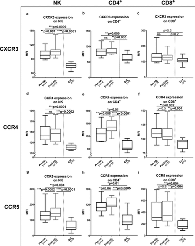
Figure 4. Different expression patterns of receptors on TILs and on PBMCs
The percentage of (a) NK cells, (b) CD4+ T cells and (c) CD8+ T cells among all lymphocytes in tumorbiopsies obtained during perfusion and in peripheral blood taken before ILP. Expression of (d-f)CXCR3, (g-i) CCR4, (j-l) CCR5 and (m-o) PD-1 on (d,g,j,m) NK cells, (e,h,k,n) CD4+ T cellsand (f,i,l,o) CD8+ T cells in tumors (TILs) and in peripheral blood (PBMCs) (n=8; paired Wilcoxon test). MFI = Median Fluorescence Intensity. Data are presented in box-and-whiskers plots with min. and max.
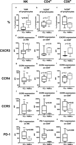
Figure 5. Melphalan-exposed melanoma cells induce expression of CXCL10, CCL2 and IFN-γ in PBMCs during co-culture
PBMCs from healthy donors were cultured with melphalan-exposed melanoma cells, non-exposed melanoma cells or were cultured alone for 48 h. After 48 h the levels of (a) CXCL10, (b) CCL2 and (c) IFN-γ were measured in the cell culture supernatants (n = 9; paired Friedman test followed by Dunn’s multiple comparison test). Data are presented as mean with SEM.
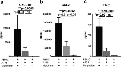
Figure 6. Melphalan-exposed melanoma cells induce expression of receptors for ISG products on PBMCs
PBMCs from healthy donors were cultured together with melphalan-exposed melanoma cells, non-exposed melanoma cells or were cultured alone. After 48 h, the PBMCs were transferred to new plates and were cultured in the absence of melanoma cells but presence of IL-2 for an additional 4 days. The expression of (a–c) CXCR3 (d-f) CCR4 (g–i) CCR5 and (j–l) PD-1 were measured on (a, d, g and j) NK cells (b, e, h and k) CD4+ T cells and (c, f, i and l) CD8+ T cells at the end of the culture (n = 6; paired Friedman test followed by Dunn’s multiple comparison test). MFI, Median Fluorescence Intensity. Data are presented as mean with SEM.
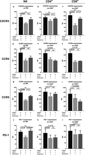
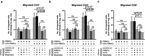
Figure 7. Activated T cells migrate toward supernatants from melphalan-exposed melanoma cells
PBMCs from healthy donors cultured for 4 days in IL-2 containing medium in the presence or absence of an anti-CD3 antibody (CD3 PBMCs; clone: OKT3) were used for chemotaxis experiments. The number of (a) CD3+, (b) CD4+ and (c) CD8+ T cells that migrated toward supernatants from PBMC-melanoma co-cultures was determined after 4 h of migration. Recombinant human CXCL10 (rhCXCL10) was used as a positive control, while medium was the negative control. (n = 6; Friedman test followed by Dunn’s multiple comparison test). The fractions of migrated T cells are presented as fold change compared to the negative control. Data are presented as mean with SEM.
Supplemental material