Figures & data
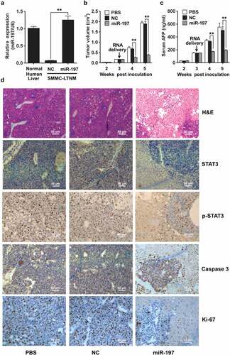
Figure 5. miR-197 suppresses HCC growth in vivo
(A) qRT-PCR analysis of miR-197 expression in normal human liver tissue, or SMMC-LTNM tumor tissue two weeks after intratumoral injection of cholesterol-conjugated miR-197 mimics (miR-197) or negative control (NC).(B–C) Two weeks after subcutaneous inoculation of SMMC-LTNM tumor cells, HCC-bearing nude mice were treated by intratumoral injection of cholesterol-conjugated miR-197, NC or PBS. Tumor volume (B), serum AFP levels (C) were shown as indicated in (C).(D) H&E staining and detection of STAT3, p-STAT3, Caspase 3 and Ki-67 by IHC in HCC tissues was performed two weeks after intratumoral injection of cholesterol-conjugated miR-197, NC or PBS. Scale bars, 50 μm.Data are shown as mean ± s.d. (n = 3) of one representative experiment. Similar results were obtained in three independent experiments. **p < 0.01.