Figures & data
Image 1. Contrast-enhanced CT images of the upper abdomen prior to initiating nivolumab (a, d) demonstrate low attenuation metastases within hepatic segments VI and II (arrows). Follow-up CT 2 years later (b, e) demonstrates that the metastasis in segment VI has decreased while the metastasis in segment II has completely resolved (arrows). Similarly, follow-up CT on 3/14/2018 (c, f) after cycle 64 of nivolumab demonstrates that the segment VI metastasis has again decreased and the segment II metastasis is still resolved (arrows)
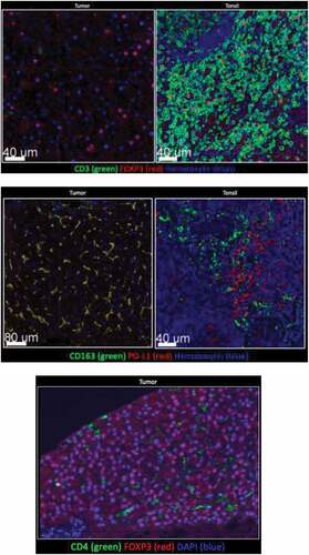
Figure 1. (continued)
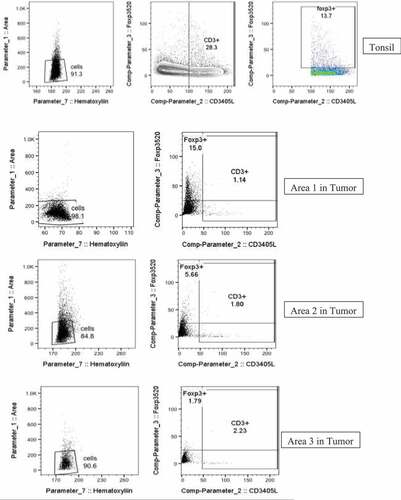
Figure 1. Tumor tissue and tonsil (control) stained for CD3, CD4, CD163, PD-L1. Few CD3+, or CD4+ cells were detected in tumor. FOXP3 was detected in tumor, but not with CD3+ or CD4+ cells. Majority of myeloid tumor cells expressed PD-L1