Figures & data
Table 1. Baseline characteristics of patients with colorectal cancer
Figure 1. Phenotypic analysis of CIK cells after expansion. (a) The phenotype of autologous CIK cells after 14-d culture from 51 patients was evaluated using flow cytometry. The positive proportions of CD3+, CD3+CD4+, CD3+CD8+, CD3−CD56+, and CD3+CD56+ are shown. (b) The phenotypic evolution of CIK cells after culture for first four treatment cycles. The results were from 49 patients and are represented as mean ± SEM. NS, not significant. * p < .05
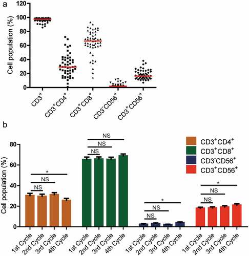
Figure 2. Kaplan–Meier estimates of disease-free survival (DFS) (a) and overall survival (OS) (b) of patients with CRC by treatment group. Significantly improved DFS and OS were observed in the CIK group (n = 60) versus the control group (n = 62)

Table 2. Univariate and multivariate analysis of disease-free survival in patients with CRC
Table 3. Univariate and multivariate analysis of overall survival in patients with CRC
Figure 3. Subgroup analysis to estimate the survival benefits from sequential CIK cell immunotherapy according to the T stage. (a) Sequential CIK cell immunotherapy slightly prolonged the disease-free survival (DFS) and overall survival (OS) of patients with T1-3 stage disease. (b) Sequential CIK cell immunotherapy significantly improved the DFS and OS of patients with T4 stage disease
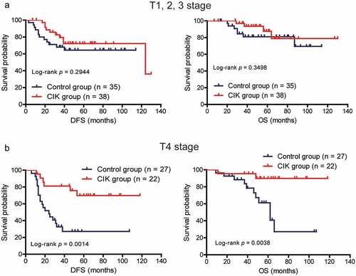
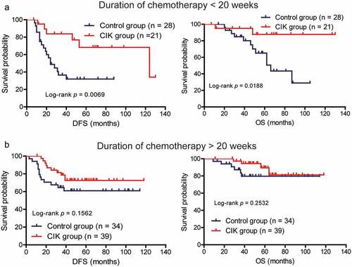
Figure 4. Subgroup analysis to estimate the survival benefits from sequential CIK cell immunotherapy according to the duration of chemotherapy. (a) Sequential CIK cell immunotherapy significantly prolonged the disease-free survival (DFS) and overall survival (OS) of patients receiving less than 20 weeks of chemotherapy. (b) Sequential CIK cell immunotherapy slightly prolonged the DFS and OS of patients receiving more than 20 weeks of chemotherapy