Figures & data
Figure 1. (a and b) Endoscopic revealed grossly normal mucosa in patients treated with anti-PD-1 and anti-CTLA-4 combination mAbs. (c) Hematoxylin and Eosin (H&E)-stained section of the colonic biopsy shows diffuse colitis with crypt atrophy and loss of crypts with residual inflamed lamina propria. A severe lymphoplasmacytic infiltration of the lamina propria with increased intraepithelial lymphocytes and crypt epithelial cell apoptosis is seen. (d and e) A severe CD4+ and CD8+ lymphocytic infiltration is seen in lamina propria. (e) Increased intraepithelial CD8+ lymphocytes (>10/100 enterocytes) and lymphocytic cryptitis are observed.
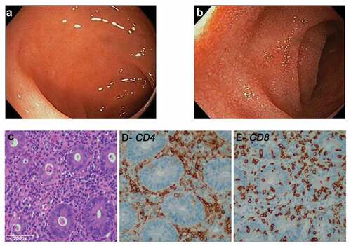
Figure 2. Treatment schema and AEs during the clinical trial. Week of cycle administration (upper line) and week at the moment of toxicity (lower line). EOT: end of treatment; FUP: follow up; G: grade; W:week; (*) Metilprednisolone.