
Figures & data
Figure 1. IL-33 causes eosinophil infiltration into tumors and reduced tumor growth in models of CRC.
(a) BALB/c mice were subcutaneously (s.c.) engrafted with 1 × 10⁵ CT26 colon cancer cells (time point 0). IL-33 treatment (0.4 µg/mouse i.p. every other day for six times) was started when tumors were palpable (after ~1 week). (b) Tumor development was monitored during the course of the treatment. Data indicate mean values ± SEM from 3 independent experiments. n = 23–25. (c) One day after the last IL-33 injection, mice were sacrificed and tumor weight and volume was measured ex vivo. Data were pooled from three independent experiments. n ≥ 29. (d) Flow cytometric analysis of single cell suspensions from s.c. tumors. Data were pooled from 3 independent experiments; n = 13–21. (e) Sirius Red staining of eosinophils in s.c. tumors of IL-33- and vehicle-treated (control) mice (representative images from n = 3/group; calibration bar: 20 µm). Arrows denote examples of Sirius Red-stained eosinophils. Inserts in the right upper corners show enlarged Sirius Red-stained eosinophils with granules easily detectable (calibration bar: 5 µm). (f) Immunoassay of lysed tumor tissue shows significant differences in the levels of IL-5 and CCL24 between IL-33- and vehicle-treated (control) mice; n = 7–10. Data indicate mean values ± SD. (g) Schematic presentation of the AOM+DSS-induced CRC model as performed in CD-1 mice treated with 1 µg IL-33/mouse i.p. 2–4 times per week. (h) Tumor number and area were measured in each colon showing a reduction in tumor growth in the AOM+DSS+IL-33 treated mice; n = 24–27. (i) Body weights (normalized to the starting weight) of IL-33- and vehicle-treated (control) mice. Data represent one of two independent experiments; n = 12–14. (j) Flow cytometric analysis of tumor infiltrated leukocytes. Data indicate mean values ± SD; n = 7–18. (k) Sirius Red staining of eosinophils in tumor tissue of AOM+DSS+IL-33- and vehicle-treated (control) mice (calibration bar: 20 µm; representative image from n = 3–4/group). Arrows denote representative examples of Sirius Red-stained eosinophils. Statistical differences were assessed by using two-way ANOVA with Sidak’s post hoc test and unpaired student’s t-test. *p < .05; **p < .01; ***p < .001.
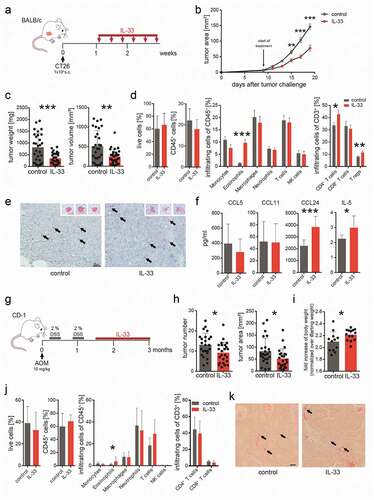
Figure 2. Expression of markers for activation, homing and degranulation.
Bone marrow-derived eosinophils were activated with IL-33 (IL-33 Eos) for 20 hrs or incubated with IL-5 (IL-5 Eos) as a control, and marker expression was evaluated using flow cytometry. (a) Histograms show representative flow cytometry experiments. Next to the histograms, the graphs show paired data of two treatments (IL-5 and IL-33). Data represent one of two independent experiments; n ≥ 5. (b) Expression of markers for homing and activation (Siglec-F and CD11b) and for degranulation (CD107a) were evaluated in tumor infiltrating eosinophils from IL-33- and vehicle-treated BALB/c mice (control) (s.c. tumor model; n = 7) and from (c) IL-33- and vehicle-treated (control) AOM+DSS mice (data pooled from two independent experiments; n = 12–18. Statistical differences were assessed by using paired and unpaired student’s t-test. *p < .05; **p < .01; ***p < .001.
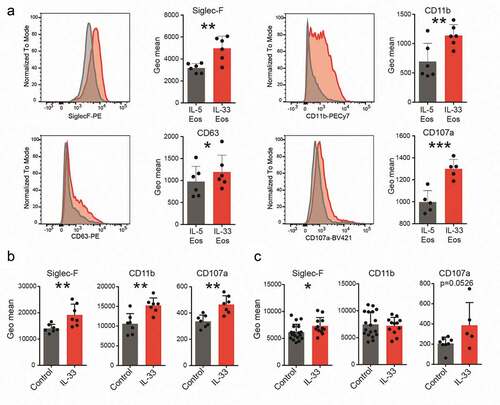
Figure 3. IL-33 dependent differences in migration and survival of eosinophils.
(a) The migration of eosinophils (pre-activated with IL-33 [IL-33 Eos] or incubated with IL-5 alone [IL-5 Eos; serving as control]) toward CT26 cell-conditioned medium and toward CCL24 is shown. Data are means ± SD, pooled from three independent experiments (n = 7) and expressed as % of vehicle (bmRPMI). (b) Eosinophil viability was measured after 24 hrs co-incubation with indicated ratios of CT26 cells and medium only, and identified as Zombie NIRTM fixable viability dye (Zombie NIR) negative cells (means ± SD, n = 3). The histogram shows a representative experiment (grey = IL-5 Eos, red = IL-33 Eos). (c) A representative flow cytometry plot shows eosinophils (IL-33 Eos and IL-5 Eos) which were injected intravenously (i.v.) into ∆dblGATA-1 mice and detected in the blood after 24 hrs. IL-33 Eos (eFluorTM 450 [eF450]-stained) and IL-5 Eos (CFSE-stained) were identified as live/CD45+/CD11b+ cells. (D, E) Graphs show IL-33 Eos and IL-5 Eos (% of CD11b+) in the blood (d) and in subcutaneous CT26 tumors 24 and 96 hrs after adoptive transfer of eosinophils into ∆dblGATA-1 mice (e); n = 4–5. Statistical differences were assessed by two-way ANOVA with Sidak’s post hoc test, multiple t-tests and by unpaired student’s t- test.*p < .05; **p < .01.
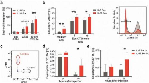
Figure 4. Eosinophils are necessary for a reduction in tumor growth by IL-33.
(a) Schematic presentation of the subcutaneous (s.c.) tumor model performed in BALB/c and eosinophil deficient ∆dblGATA-1 mice. IL-33 was given i.p. at a concentration of 0.4 µg/mouse every other day for a total of six times. (b) Tumor growth was monitored during the course of the experiment; n ≥ 8. Data indicate mean values ± SEM. **p < .01 BALB/c + control vs. BALB/c + IL-33; no significance between other groups. (c) One day after the last IL-33 injection, BALB/c and ∆dblGATA-1 mice were sacrificed and tumor weight and volume were measured ex vivo showing no differences between IL-33- and vehicle (control) treatment in the ∆dblGATA-1 but tumor growth reduction in the BALB/c mice; n ≥ 23, three independent experiments. (d) Flow cytometric analysis of single cell suspensions of tumors from BALB/c and ∆dblGATA-1 mice indicating significant differences between IL-33- and vehicle-treated (control) ∆dblGATA-1 mice for CD45+, monocytes, CD8+ T cells and Tregs, as well for eosinophils between IL-33- and vehicle-treated (control) BALB/c mice; n = 24; three independent experiments. (e) ∆dblGATA-1 mice with s.c. tumors were repopulated by adoptive transfer (twice weekly) with either IL-33 Eos or IL-5 Eos (eosinophils were isolated from bone marrow of BALB/c mice and differentiated). (f) Adoptive transfer with IL-5 Eos failed to significantly restore tumor reduction in ∆dblGATA-1 mice (in comparison to ∆dblGATA-1 mice given PBS only). (g) However, adoptive transfer with IL-33 Eos (vs. PBS only) significantly restored reduction of tumor growth; n = 8–9. (h) Single cell suspensions of the s.c. tumors were prepared and infiltrated eosinophils were evaluated as % of CD11b+ cells; n = 8–15. (i) Immunohistochemistry of s.c. tumors with an anti-EPX antibody shows that PBS-treated ∆dblGATA-1 mice are devoid of eosinophils. After adoptive transfer of IL-33 Eos (by i.v. injection), EPX staining is visible in the tumors of ∆dblGATA-1 mice. Infiltrated eosinophils in s.c. tumors of BALB/c mice are shown for comparison (representative images of n = 3/group; calibration bar: 20 µm). Statistical differences were assessed by using multiple t-tests, one-way ANOVA with Tukey’s multiple comparisons test and unpaired student’s t-test. *p < .05; **p < .01; ***p < .001, ns = not significant.

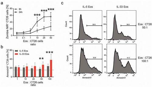
Figure 5. Cytotoxicity of eosinophils against CT26 cells in vitro.
(a) Bone marrow-derived eosinophils (Eos) were co-incubated with CT26 cells at different eosinophil (Eos):CT26 cell ratios for 6 and 24 hrs. The percentage (%) of dead (Zombie NIR+) CT26 cells is depicted as mean ± SD; n = 5. (b) CT26 cells were co-incubated with either IL-5 Eos or IL-33 Eos at different Eos:CT26 cells ratios for 7 hrs (n = 5). Percentages (%) of AnnexinV+ CT26 cells are shown. (c) Representative histograms of IL-5 Eos and IL-33 Eos co-incubated with CT26 cells for 7 hrs at indicated ratios. Statistical differences were assessed using two-way ANOVA with Sidak’s post hoc test. **p < .01, ***p < .001.
Supplemental material