Figures & data
Table 1. Baseline characteristics of patients.
Figure 1. Reduction in Tregs and dynamics of ERK phosphorylation on c-Kit+Tregs during tivozanib therapy (A) Representative histogram offset showing frequency of CD4+Foxp3+ Tregs measured at pre, 28–35d and 160d of tivozanib treatment. (B) Frequency and (C) absolute numbers of CD4+Foxp3+ Tregs pre vs 28–35d (D) Representative histogram offset showing frequency of Foxp3+c-Kit+ Tregs measured at pre, 28–35d and 160d of tivozanib treatment. (E) Frequency and (F) absolute numbers of Foxp3+c-Kit+ Tregs pre vs 28–35d (G) Representative histogram offset showing frequency of c-Kit+pPERK+ Tregs measured at pre, 28–35d and 160d of tivozanib treatment. (H) Frequency and (I) absolute numbers of c-Kit+pPERK+ Tregs pre vs 28–35d (J) Ratio of CD4+CD127+ T cells to CD4+Foxp3+ T cells (CD4+CD127+ T cells/CD4+Foxp3+ T cells) pre vs 28–35d. (K) Ratio of CD8+CD127+ T cells to CD4+Foxp3+ T cells pre vs 28–35d. Each symbol represents an individual HCC patient. Frequencies of Tregs and T effector cells were calculated based on CD3+CD4+ T/CD3+CD8+ T cell population. **** P < 0.0001, *** P < 0. 001, ** P < 0.01, * P < 0.05, paired t-test, Pre vs 28–35d n = 17.
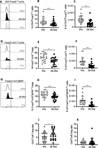
Figure 2. Reduction in MDSC and dynamics of ERK phosphorylation on c-Kit+ MDSCs during tivozanib therapy. (A) Representative histogram offset showing frequency of CD11b+CD33+ MDSCs measured at pre, 28–35d and 160d of tivozanib treatment. (B) Frequency and (C) absolute numbers of MDSCs pre vs 28–35. (D) Representative histogram offset showing frequency of c-Kit+ MDSCs measured at pre, 28–35d and 160d of tivozanib treatment. (E) Frequency and (F) absolute numbers of c-Kit+ MDSCs pre vs 28–35. (G) Representative histogram offset showing frequency of c-Kit+pERK+ MDSCs measured at pre, 28–35d and 160d of tivozanib treatment. (H) Frequency and (I) absolute numbers of c-Kit+pERK+ MDSCs pre vs 28–35. Each symbol represents an individual HCC patient. Frequencies of MDSCs were calculated based on CD14−HLA-DR− population **** P < 0.0001, *** P < 0. 001, ** P < 0.01, * P < 0.05, paired t-test, n = 17.
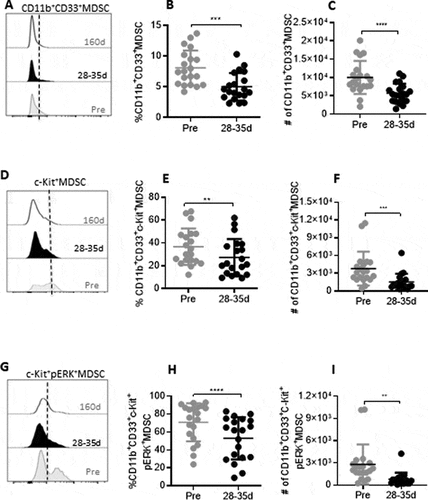
Figure 3. Reduction in immune checkpoint receptors during and after tivozanib therapy. (A) Representative histogram offset showing frequency of Foxp3+CTLA-4+ Tregs measured at pre, 28–35d and 160d of tivozanib treatment. (B) Frequency and (C) absolute numbers of Foxp3+CTLA-4+ Tregs pre vs 28–35d. (D) Representative histogram offset showing frequency of CD4+PD-1+ T cells measured at pre, 28–35d and 160d of tivozanib treatment. (E) Frequency of CD4+PD-1+ T cells pre vs 28–35d (F) Absolute number of CD4+ PD-1+ T cells pre vs 28–35d (G) Representative histogram offset showing frequency of CD8+PD-1+ T cells measured at pre, 28–35d and 160d of tivozanib treatment. (H) Frequency of CD8+PD-1+ T cells pre vs 28–35d (I) Absolute number of CD8+PD-1+ T cells pre vs 28–35d. Each symbol represents an individual HCC patient. Frequencies of CTLA-4+Tregs and PD-1+ T cells were calculated based on CD3+CD4+ T cell/CD3+CD8+ T cell population and. **** P < 0.0001, *** P < 0. 001, ** P < 0.01, * P < 0.05, paired t-test, n = 17.
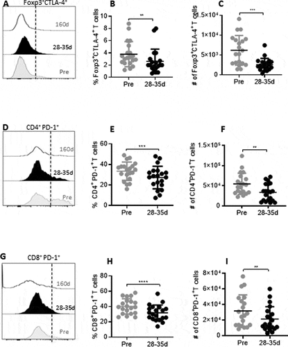
Figure 4. Kaplan-Meier plots showing the predictive immune correlates of responses to tivozanib treatment in HCC patient. Association between immunophenotypic signatures and OS or PFS of patients was calculated as described in methods. (A) OS of the patients and increased frequencies of baseline CD4+PD-1+ T cells (HR = 0.92, 95% CI: 0.9–1.0, P = 0.02). (B) Reduction in the frequencies of CD4+Foxp3+ Tregs (post- pre changes) (median reduction: −2.5, range: −7.6 to −0.8), quantified after tivozanib therapy and OS of the patients (HR = 1.6, 95% CI: 1.0–2.3, P = 0.03). (C) Low baseline ratio of CD4+T effector cells to Foxp3+ Tregs and OS of the patients (HR = 1.2, 95% CI: 1.0–1.5, P = 0.046) as well as (D) Low baseline ratio of CD4+T effector cells to Foxp3+ Tregs and PFS of the patients at 24 weeks (HR = 1.4, 95% CI: 1.0–1.9, P = 0.03). (E). Decrease in the frequencies of Foxp3+c-Kit+pERK+Tregs (post-pre changes) and PFS of the patients at 24 weeks (HR = 1.2, 95% CI: 1.0–1.5, P = 0.03).
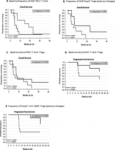
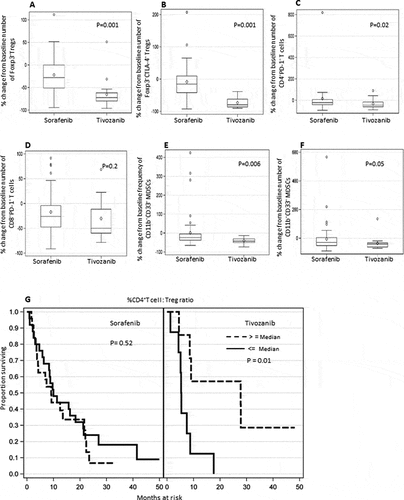
Figure 5. Differential effect of tivozanib vs sorafenib treatment on immune suppressive cell subsets in HCC patients and impact of post-treatment changes in the ratio of CD4+T effector cell: Foxp3+ Tregs on overall survival of HCC patients after tivozanib vs sorafenib treatment. Box plots represent mean/median percentage changes from baseline number or frequency of different immune cell subsets after 28–25 days of sorafenib vs. tivozanib treatment (A) Foxp3+ Tregs (B) Foxp3+CTLA-4+ Tregs (C) CD4+PD-1+ T cells (D) CD8+PD-1+ T cells (E) % CD11b+CD33+ MDSC (F) # CD11b+CD33+ MDSC (G) Kaplan-Meir plots showing the association of greater percentage change or increase in the ratio of CD4+T cells: Tregs from baseline and survival probability of HCC patients after sorafenib or tivozanib treatment. P values are shown inside the respective plots; sorafenib n = 49, tivozanib n = 17.