
Figures & data
Figure 1. Neutrophils resistant to anti-Ly6G depletion exhibit ANCAs
(a). Ly6G binding and trafficking: untreated BM cells were incubated with DMEM or cytochalasin D before anti-Ly6G-PE staining. After 1 hour of incubation, bone marrow (BM) cells were stained with CD45-APC. (b). Representative images obtained with imagestream to localize Ly6G-PE. (c.d). Quantification from b (n = 3 DMEM, n = 4 Cytochalasin D, 500 neutrophils/sample). (e) Gated on immune cells. Sensitive strategy based on S100A9 and intra-cellular Ly6G staining to detect neutrophils after anti-Ly6G treatment. Note that only the MFI for intra-cellular Ly6G is lower in anti-Ly6G treated mice. f Neutrophil prevalence quantification in BM, blood and lungs after 5 d of isotype CTR or anti-Ly6G treatment (n = 5 mice per group, 25 µg of ab, daily). g. Scheme of experiment to track anti-Ly6G delivered in-vivo after 5 d of treatment (25 µg of ab, daily). Excess unlabeled anti-Ly6G (200 ml at 10µ g/mL for 15 minutes) is added prior fixation to allow CTR cell detection with an anti-rat-FITC. h. Imagestream images obtained from g are compatible with anti-neutrophil cytoplasmic antibodies (ANCAs) in BM neutrophils. Neutrophils were gated on the basis of the FITC positive signal: note that both anti-rat antibodies are polyclonal and displayed no competition at the surface membrane. DAPI stained nucleus shows several segmentations, which confirms the granulocytic nature of gated cells. (i) Quantification of the intracellular presence of anti-Ly6G ab after 5 d of treatment in the BM, or 1 hour of treatment in the blood (the blood endpoint was added from an additional experiment).j Representative illustration on projected BM cells showing the in-vivo delivered anti-Ly6G colocalizes with calnexin, an endoplasmic reticulum protein).* p < .05, ** p < .01, *** p < 0,001, **** p < 0,0001 from Mann-Whitney test; error bars represent s.d.
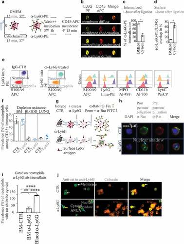
Figure 2. Anti-Ly6G ligation enhances TNFα-induced neutrophil oxidative burst
(a-b). Five mice were treated with anti-Ly6G or isotype CTR for 5 d. Bone marrow (BM) and blood cells were collected and incubated with or without TNFα for 1 hour at 37°C. Cells were subsequently stained with the pauci-competitive anti-Gr1at the surface a and Ly6C (b). Note that the surface levels for Gr1 in the blood neutrophils are lower than levels in the BM, while the neutrophil exit in physiological conditions normally goes with an increase of surface Ly6G, suggesting an active loss of the Ly6G antigen upon in vivo anti-Ly6G treatment p < .05, ** p < .01, *** p < 0,001, **** p < 0,0001 from Mann-Whitney test; error bars represent s.d. (c). Schematic representation of the Ly6G membrane availability upon anti-Ly6G after treatment induction and maintenance. (d). 200000 BM cells were incubated with anti-Ly6G for 15 minutes at 4°C (or isotype CTR, 10 µg/mL), extensively washed, pulsed with TNFα (5 ng/mL) for 30 min at 37°C, and incubated with a ROS detecting probe for 0–15-30 or 60 minutes (n = 6 per group per endpoint). **** p < 0,0001 from 2way ANOVA test; error bars represent s.d e. The experimental setting from d was reproduced for the endpoint 15 min, in the presence of PBS, FcBlock or Cytochalasin D to evaluate the role of residual surface Fc-FcGr interactions as well as the internalization of the Ly6G/anti-Ly6G complex on the enhanced ROS production. Six bone marrows coming from six untreated mice were used. Each bone marrow is divided to test all conditions. treatment p < .05, ** p < .01, *** p < 0,001, **** p < 0,0001 from ANOVA test with multiple comparison; error bars represent s.d
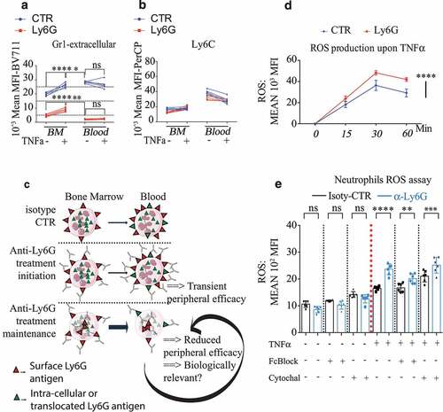
Figure 3. TAN aging enables SiglecF differentiation and depletion resistant Ly6Gneg neutrophils
(a). Scheme of treatment: KP mice received 1 mg of BrdU injected i.p (single dose) and sacrificed at dedicated endpoints to correlate neutrophil aging with SiglecF expression. (b). representative FACS plot gated on CD45+ CD11b+Ly6GhighF4/80neg SSCalow TANs at each endpoint. (c). SiglecF proportion among all TANs in terminal KP tumor tumors. (Merge of three independent experiments labeled in different colors) (d). TAN BrdU+ fraction according to the SiglecF status from the arrival of BrdU+ TANs 60 h post-BrdU injection, to their extinction 240 post-BrdU injection. Each dot is a quantification of a single tumor. Each endpoint is a single experiment with 4 KP mice per group, 6 to 12 micro-dissected single tumors analyzed. **** p < 0,0001 from one-way ANOVA; error bars represent s.d. (e-f). Representative dot plot, gated on CD45+ CD11b+ immune cells, from KP treated for 8 d with anti-Ly6G or relevant isotype CTR. After anti-Ly6G, anti-Ly6G is sensitive to detect S100A9 neutrophils in lungs but not in tumors. (g). Estimation of false negatives upon treatment in old SiglecFpos TANs. **** p < 0,0001 from Mann-Whitney test; error bars represent s.d. (h). Differential ly6g expression at the transcriptomic level (data extrapolated from an online single-cell RNA database).
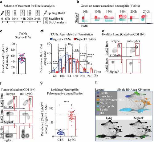
Figure 4. Neutrophil depletion alone has no anti-tumor effect
(a). Experimental design: KP-tumor-bearing mice were exposed for 2 weeks with different treatment regimen affecting neutrophil. Top: anti-Ly6G1A8, anti-Gr1-RB6 were injected i.p every other day (100 µg/mice). G-CSF is injected daily subcutaneously (7,5 µg/mice). Bottom: combination of anti-Ly6G+anti-rat abs to incrementally optimize the killing effect of anti-Ly6G and be comparable to anti-Gr1. Tumor follow-up is performed by CB-CT scan at d0 and d14. Each tumor is contoured using the medical Osirix software and a 3D volume reconstitution is performed. (b): relative to D0 tumor growth after 14 d of treatment. Results obtained from 3 to 7 KP mice per group; (c): TAN prevalence at the end of treatment completion. (d). relative to D0 tumor growth after 14 d of treatment. results obtained from 7 KP mice per group. e.Anti-Ly6G+antirat combination has no anti-tumor effect despite it reduces TANs by >50%. Results from b + c and d + e comes from independent experiment. * p < .05, ** p < .01, *** p < 0,001, **** p < 0,0001 from Mann-Whitney test; error bars represent s.d.
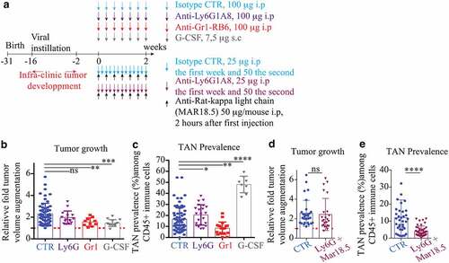
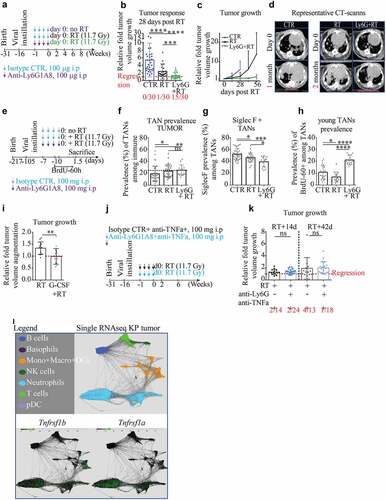
Figure 5. Anti-Ly6G enhances the recruitment of young neutrophils and synergizes with RT
(a). Scheme of treatment: well-established KP tumor-bearing mice received 1 week of isotype/anti-Ly6G ab (100 µg/mice, once every other day), followed by a radiation (RT) treatment at day 0. (b.c). Tumor response is measured as relative growth compared to day 0 by CB-CT scan. Five mice per groups. The number of regressive tumors is given below and indicates regressing tumors. Because untreated mice systematically give rise to escapers and tumor merge, the follow-up had to be stopped after 1 month in CTR group. (d). Illustrative CB-CT scan-based follow up at indicated endpoints. €. Experimental design to assess early changes after treatment termination in single micro-dissected tumors (f.g.h), each dot is a single micro-dissected tumor). f. TAN prevalence among all immune cells. g. Prevalence of old SiglecF+ TANs among all TANs. h. Prevalence of “young” TANs of 60 hours or less (arrival phase). (i). Because the radiosensitizing effect of anti-Ly6G is associated with an increase of young infiltrating TANs, it suggests a potential gain of function of residual neutrophils. KP mice received a single dose of 7.5 µg of G-CSF one-hour prior RT and tumor growth was assessed as in a. j. Scheme of treatment: well-established KP tumor-bearing mice receive 1 week of anti-TNFα ± anti-Ly6G prior to RT (11.7 Gy). k. 2- and 6-weeks post-RT relative tumor growth: the radiosensitizing effect of anti-Ly6G is not recapitulated in the presence of anti-TNFα. i. Expression of Tnfrsf1b and Tnfrsf1a in KP-TAN (data extrapolated from single-cell RNA sequencing available online). Data from b-c; f-h; i and k are derived from independent experiments with 3 to 7 KP mice per groups. Each dot represents a single tumor. * p < .05, ** p < .01, *** p < 0,001, **** p < 0,0001 from Mann-Whitney test; error bars represent s.d.
Supplemental material