
Figures & data
Figure 1. utse outgrowth over time. Schematic of utse outgrowth over time (A) Early L4 vulva and uterus. The utse has just formed at this stage after the fusion of eight ρ cells and the anchor cell. The cell has an ellipsoidal shape. Outlines indicate positions of vulva and uterus. utse is indicated in red. Blue dashed arrows indicate direction of outgrowth. (B) Mid L4 vulva and uterus. utse has begun elongating along the anterior-posterior axis. Outlines indicate positions of vulva and uterus. utse is indicated in red. (C) L4 lethargus vulva and uterus. utse has completed its outgrowth, and has taken on an elongated shape with the edges of its arms extending along the dorsal/ventral axis. Outlines indicate positions of vulva and uterus. utse is indicated in red.
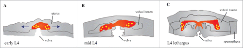
Figure 2. Vulval sex muscle outgrowth. Schematic of a sex myoblast at the L3 larval stage. A cross section of an L3 vulva shows that a sex myoblast daughter cell (in red) is in between the vulval epithelium and the seam cells of the hypodermis. Sex myoblasts extend protrusions ventrally towards the vulva and laterally towards the seam cells.
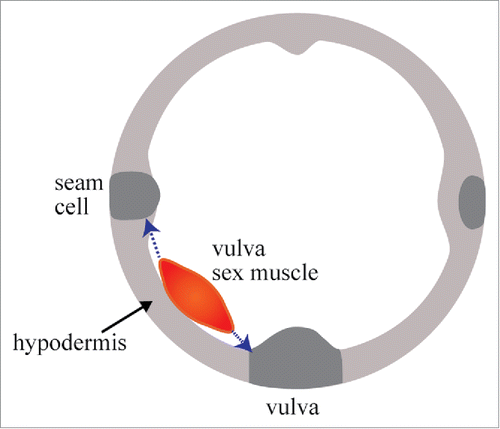
Figure 3. Anchor cell invasion. Schematic anchor cell invasion. Anchor cell shown in red, vulva shown in gray. Basement membrane shown in thick line. Adapted from Ihara et al., 2011 and Hagedorn and Sherwood, 2011. (A) Anchor cell at P6.p two cell stage during mid L3. Basement membrane (shown in thick line) is intact. Anchor cell is dorsal to the P6.p 1° VPC daughters (two circles below). (B) Anchor cell at P6.p four cell stage during mid to late L3. Anchor cell has generated a gap in the basement membrane (see separation between thick lines). Edges of the anchor cell are still in contact with the basement membrane. (C) Anchor cell at P6.p late four-cell stage (late L3). The anchor cell has begun forming protrusions that will invade the vulva (dashed purple arrow). (D) Anchor cell at P6.p late four-cell stage (late L3). The anchor cell has completely invaded the vulva, specifically invading between the 1° VPC granddaughters. (E) Anchor cell at early L4 stage. The 1◦ VPCs have divided and proximal cells are shown. Vulval invagination has occurred and anchor cell will soon fuse with the ρ cells to form the utse.
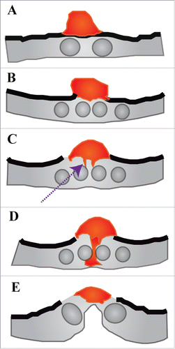
Figure 4. Muscle arm extension. Schematic of muscle arm extension. Adapted from Dixon and Roy, 2005. Example of muscle arms throughout the C. elegans body (light blue arrows). Muscle arms have formed from the dorsal body wall muscles (end of dark blue arrow) towards the dorsal nerve cord.
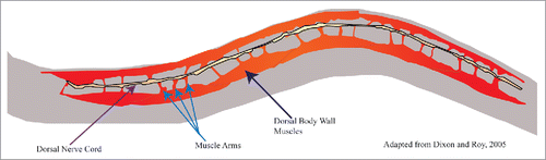
Figure 5. Male tail cell shape change and outgrowth. Schematic of male tail retraction and ray formation from L3 to adulthood. Adapted from Nguyen et al., 1999. (A) Male tail at L3 stage, retraction and ray formation have not occurred and entire cell is composed of tail epithelium. (B) Beginning of tail retraction in L4. Light red indicates fluid-filled extracellular space previously inhabited by tail epithelium. (C) L4 stage. Continuation of retraction in male tail, start of ray formation. (D) L4 stage, male tail has finished retracting and rays have all formed. (E) Adult male tail. Rays have reached their final shape. Fluid-filled extracellular space has taken peloderan shape.
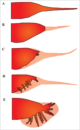
Figure 6. Excretory cell outgrowth. Schematic of excretory cell outgrowth. Excretory cell in red, outline of worm in black. Adapted from Buechner et al., 2002. (A-D) Excretory cell outgrowth during three-fold embryonic stage. (A) Excretory cell at birth, cell has a spherical shape. (B) Cell takes on an ellipsoidal shape as it begins to grow laterally from left to right over the ventral muscle quadrants. (C) Apical and basal edges of the excretory cell begin migrating dorsally. (D) Edges of dorsal protrusions bifurcate and begin to grow outward laterally along the anterior posterior axis. (E) Completed excretory cell outgrowth (L1). Cell has extended laterally along the anterior-posterior axis to span the entire length of the worm.
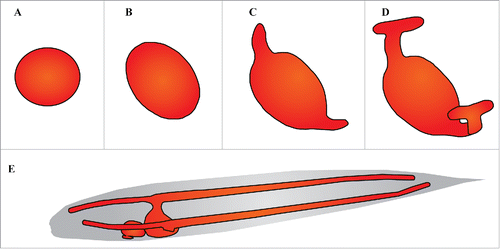
Figure 7. Head mesodermal cell. Schematic of head mesodermal cell. Positioning of head mesodermal cell in adult worm. Head mesodermal cell lies dorsomedial to the terminal bulb of the pharynx. It extends processes that split at the pharynx and extend anteriorly and posteriorly along the dorsal and ventral margins of the body wall. These processes also lie adjacent to the intestine as well as the excretory gland cell. Adapted from Altun and Hall, 2009.
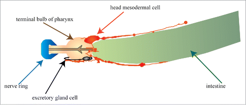
Figure 8. Dorsal intercalation. Schematic of dorsal intercalation. Adapted from Chisholm and Hardin, 2005. (A) Dorsal view of embryo undergoing intercalation. Intercalating epidermal cells shown in red. Dashed lines indicate cells that have already intercalated. Cells that are intercalating are changing from a rounded shape to a more wedge/protrusion-like shape. (B) Expanded view of intercalating cells. Basolateral protrusions that also touch neighboring cells, and help the intercalating cells move towards one another.
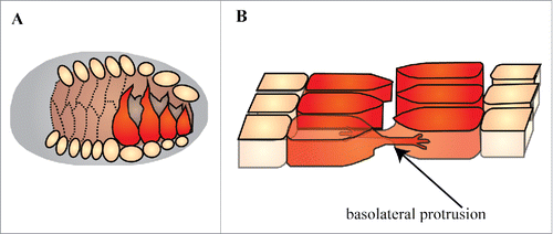
Figure 9. Ventral Enclosure. Schematic of ventral enclosure. Adapted from Chisholm and Hardin, 2005. Ventral epidermal cells shown in red, neuroblasts shown in gray spheres. (A) Outgrowth of epidermal cells during ventral enclosure. Ventral marginal cells are indicated with blue arrow and pocket cells are indicated with green arrow. (B) Expanded view of ventral epidermal cells moving along the neuroblast substratum.
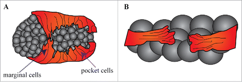
Table 1. Genes involved in cell outhgrowth.
Table 2. Characteristics used by different outgrowth systems.