Figures & data
Figure 1. Vaccination and rotometry testing schedule and amino acid sequences of DC sensitizing peptides. (A) Schedule for vaccination, rotometry testing and euthanasia as described further in the Materials and Methods section. The initial vaccination with sensitized or non-sensitized DCs was made in 3.5 month old α− Syn expressing Tg mice. (B) Amino acid sequence and residue numbers for the 3 α-Syn specific B cell epitope-containing peptides used as DC sensitizers.
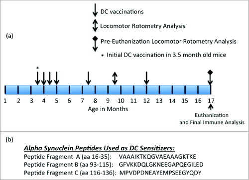
Figure 2. Anti-α− Syn antibody responses elicited by PSDC or rh- α− Syn or rh- sensitized DC vaccines as measured by ELISA. (A) Ten days following the initial vaccination PSDC administered Tg (i.e. Tg PSDC) mice demonstrated a significantly higher anti-α−Syn antibody response, measured by OD450nm values, than did Tg mice vaccinated with rh-α− Syn sensitized DCs (i.e., Tg rh-α− Syn). OD450nm binding values are also provided for non-sensitized PSDC vaccinated mice (i.e. Tg DC Control). (B) Time course of anti-α− Syn antibody responses from analysis of sera from either PSDC (i.e., Tg PSDC) or rh-α− Syn DC sensitized (i.e. Tg rh-α− Syn DC) vaccinated mice. OD450nm binding values are also provided for non-sensitized DC vaccinated mice (i.e., Tg DC Control) (C) Demonstration that sensitization of DCs with peptide fragment C resulted in the highest anti-α− Syn peptide antibody responses of the 3 peptides tested. For analysis of data presented in (A), (B) and (C) above statistical significance was determined by the student t test with differences being significant at the 0.05 level and are indicated by *.
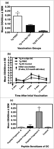
Figure 3. Rotometric locomotor performance of Tg mice at age 17 months after the vaccination regimen with either PSDC or rh-α− Syn sensitized DCs. The PSDC (i.e. Tg PSDC) or rh-α− Syn sensitized (i.e., Tg rh-α− Syn) DC vaccinated Tg mouse groups performed significantly better (i.e. higher latency values) on the rotorod test than did Tg mice vaccinated with non-sensitized DCs (i.e., Tg DC Control). Latency values are also provided for WT (wild type) control mice (i.e. WT Control).
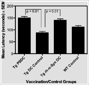
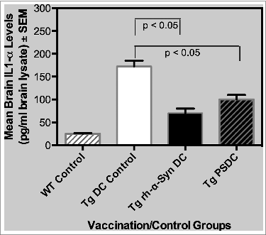
Figure 4. Levels of the pro-inflammatory cytokine IL-1α in brain lysates from PSDC or rh-α− Syn sensitized DC vaccinated mice. Levels of IL-1α in brain lysates are expressed as pg/ml ± SEM. The results indicate that significantly lower levels of IL-1α were measured in the brain lysates from PSDC or rh-α− Syn sensitized DC vaccinated Tg mice than in non-sensitized DC vaccinated Tg controls. IL-1α levels are also provided for WT (wild type) control mice (i.e., WT Control).