Figures & data
Figure 1. Characterization of anti-Tat humoral responses in sera. Serum samples of mice immunized 3 times ID with Tat protein at the dose of 1, 7.5 or 30μg were collected at day 42 from retro-orbital plexus and the presence of anti-Tat IgG and IgM was evaluated by Elisa test. Briefly, 96-well plates were coated with Tat (100ng/200μl/well) in 0.05 M carbonate buffer (pH 9.6) for 18 hours at 4°C. Plates were washed with PBS containing 0.05% Tween 20 (Sigma-Aldrich, Milan, Italy), and incubated for 90 minutes at 37°C with blocking buffer. The following blocking buffers were used: PBS containing 0.05% Tween 20 and 1% BSA (for IgG) and PBS containing 0.05% Tween 20 and 3% BSA (for IgM). After extensive washes, serial dilutions of each serum were dispensed in duplicate wells (100μl/well) and incubated for 90 minutes at 37°C. Plates were washed again before the addition of 100 μl/well of HRP-conjugated goat anti-mouse IgG or IgM (Sigma), and incubated at 37°C for 90 minutes. After incubation, plates were washed 5 times and subsequently a solution of 2,2'-Azinobis [3-ethylbenzothiazoline-6-sulfonic acid]-diammonium salt (ABTS) substrate (Roche) was added. The absorbance values were measured at 405 nm with an automatic plate reader (SUNRISE TECAN Salzburg-Austria). The cut-off value was estimated as the mean OD of 3 negative control sera plus 0.05. Each OD value was subtracted of the blank and cut-off values to obtain a net OD value and IgG titers calculated by intercept function. (A) Titers of serum anti-Tat IgG. (B) Titers of serum anti-Tat IgM. *P < 0.05, **P < 0.01 according to 2-tailed Mann Whitney test. Results of 2 independent experiments are shown. Dots represent single mice and lines represent the means +/− SEM.
![Figure 1. Characterization of anti-Tat humoral responses in sera. Serum samples of mice immunized 3 times ID with Tat protein at the dose of 1, 7.5 or 30μg were collected at day 42 from retro-orbital plexus and the presence of anti-Tat IgG and IgM was evaluated by Elisa test. Briefly, 96-well plates were coated with Tat (100ng/200μl/well) in 0.05 M carbonate buffer (pH 9.6) for 18 hours at 4°C. Plates were washed with PBS containing 0.05% Tween 20 (Sigma-Aldrich, Milan, Italy), and incubated for 90 minutes at 37°C with blocking buffer. The following blocking buffers were used: PBS containing 0.05% Tween 20 and 1% BSA (for IgG) and PBS containing 0.05% Tween 20 and 3% BSA (for IgM). After extensive washes, serial dilutions of each serum were dispensed in duplicate wells (100μl/well) and incubated for 90 minutes at 37°C. Plates were washed again before the addition of 100 μl/well of HRP-conjugated goat anti-mouse IgG or IgM (Sigma), and incubated at 37°C for 90 minutes. After incubation, plates were washed 5 times and subsequently a solution of 2,2'-Azinobis [3-ethylbenzothiazoline-6-sulfonic acid]-diammonium salt (ABTS) substrate (Roche) was added. The absorbance values were measured at 405 nm with an automatic plate reader (SUNRISE TECAN Salzburg-Austria). The cut-off value was estimated as the mean OD of 3 negative control sera plus 0.05. Each OD value was subtracted of the blank and cut-off values to obtain a net OD value and IgG titers calculated by intercept function. (A) Titers of serum anti-Tat IgG. (B) Titers of serum anti-Tat IgM. *P < 0.05, **P < 0.01 according to 2-tailed Mann Whitney test. Results of 2 independent experiments are shown. Dots represent single mice and lines represent the means +/− SEM.](/cms/asset/aea08b41-7e69-485b-9139-fe91ed6e64cf/khvi_a_1016676_f0001_b.gif)
Figure 2. Characterization of anti-Tat humoral responses in sera. Serum samples of mice immunized 3 times ID, IM or OM with Tat protein (30μg) were collected at day 42 and the presence of anti-Tat IgG, IgM and IgA was evaluated by Elisa test. Elisa test for IgA evaluation was performed using PBS containing 0.1% Tween 20 and 1% BSA as block buffer. (A) Proportion of mice that developed serum anti-Tat IgG, IgM and IgA. Frequencies of anti-Tat positive mice were compared among different groups using 2-tailed Fisher's exact test. Anti-Tat positivity was determined by titers >100, 50 and 25 for IgG, IgM and IgA respectively. (B) Titers of serum anti-Tat IgG. (C) Titers of serum anti-Tat IgM. (D) Titers of serum anti-Tat IgA. * P < 0.05, **P < 0.01 according to 2-tailed Mann Whitney test. Results of 2 independent experiments are shown. Dots represent single mice and lines represent the means +/− SEM.
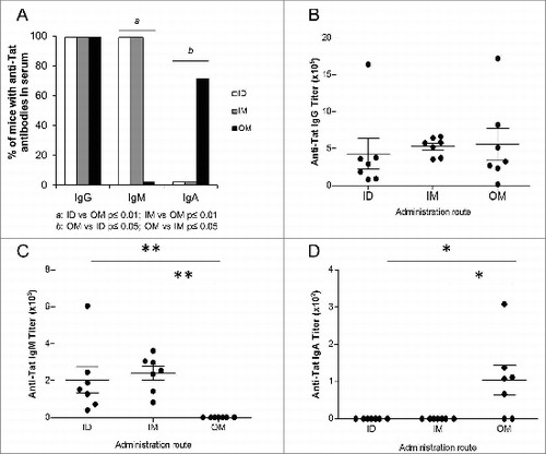
Figure 3. Characterization of anti-Tat humoral responses in mucosal lavages. Mucosal samples of mice immunized 3 times ID, IM or OM with Tat protein (30μg) were obtained at day 42 by repeated flushing and aspiration with 0.5 ml of PBS containing a protease inhibitor cocktail (Roche, Mannheim, Germany). The presence of anti-Tat IgG and IgA was evaluated by Elisa test. Proportion of mice that developed anti-Tat IgG and IgA in (A) vaginal and (D) intestinal lavages. Frequencies of anti-Tat positive mice were compared among different groups using 2-tailed Fisher's exact test. Anti-Tat positivity was determined by titers >50 and 25 for IgG and IgA respectively. Titers of anti-Tat IgG in (B) vaginal and (E) intestinal lavages. Titers of anti-Tat IgA in (C) vaginal and (F) intestinal lavages. * P < 0.05 according to 2-tailed Mann Whitney test. Results of 2 independent experiments are shown. Dots represent single mice and lines represent the means +/− SEM.
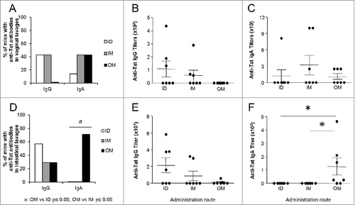
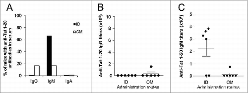
Figure 4. Characterization of anti-Tat 1–20 peptide humoral responses in sera. Serum samples of mice immunized 3 times ID or OM with the Tat 1–20 peptide (7μg) were collected at day 42 and the presence of anti-Tat 1–20 peptide IgG, IgM and IgA was evaluated by Elisa test (plates were coated with Tat 1–20 peptide at the dose of 100ng/200μl/well). (A) Proportion of mice that developed serum anti-Tat 1–20 peptide IgG, IgM and IgA. Frequencies of anti-Tat 1–20 peptide positive mice were compared among different groups using 2-tailed Fisher's exact test. Anti-Tat 1–20 peptide positivity was determined by titers >100, 50 and 25 for IgG, IgM and IgA respectively. (B) Titers of serum anti-Tat 1–20 peptide IgG. (C) Titers of serum anti-Tat 1–20 peptide IgM. Results of 2 independent experiments are shown. Dots represent single mice and lines represent the means +/− SEM.