1,290
Views
13
CrossRef citations to date
0
Altmetric
Research Paper

Presence of mycobacterial L-forms in human blood: Challenge of BCG vaccination

, &
Pages 1192-1200 | Received 15 Dec 2014, Accepted 23 Jan 2015, Published online: 27 May 2015

Figures & data

Figure 1. “Non-cultivable elements” from blood isolates (No1; 34; 37, 48; 91; 96). Light microscopy of: (A, B, C) spherical and filamentous forms found in semisolid agar during the first phase/week of cultivation; (D, E, F) initiation of growth from filamentous elements during the second phase/week of cultivation. Magnification: 200x
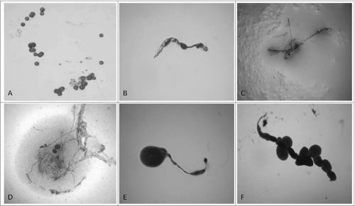
Figure 1. “Non-cultivable elements” from blood isolates (No1; 34; 37, 48; 91; 96). Light microscopy of: (A, B, C) spherical and filamentous forms found in semisolid agar during the first phase/week of cultivation; (D, E, F) initiation of growth from filamentous elements during the second phase/week of cultivation. Magnification: 200x

Figure 2. Development of L-form growths in semisolid agar during the second phase/week of cultivation. Light microscopy of blood isolates (No1; 34; 37, 48; 91; 96): typical “fried eggs” colonies of different size, consistence and density (A, B, C, D); formation of biofilm with gliding motility at the periphery (E, F). Magnification: 400x
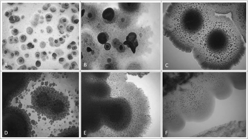
Figure 2. Development of L-form growths in semisolid agar during the second phase/week of cultivation. Light microscopy of blood isolates (No1; 34; 37, 48; 91; 96): typical “fried eggs” colonies of different size, consistence and density (A, B, C, D); formation of biofilm with gliding motility at the periphery (E, F). Magnification: 400x

Figure 3. SEM of huge filamentous and membranous L-type formations with many small granular, oval or coccoid cells fit together (A-E) and tightly packed rods (F) from blood isolate No1 grown in semisolid agar during the second phase/week of cultivation. Bars: 100 μm (D); 10 μm (A, B, E, F).
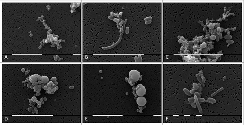
Figure 3. SEM of huge filamentous and membranous L-type formations with many small granular, oval or coccoid cells fit together (A-E) and tightly packed rods (F) from blood isolate No1 grown in semisolid agar during the second phase/week of cultivation. Bars: 100 μm (D); 10 μm (A, B, E, F).

Figure 4. SEM of L-form cells from blood isolate No1 grown in semisolid agar during the second phase/week of cultivation. (A) Cluster of granular forms; (B, C, F) groups of polymorphic granular, oval, rod and elongated filamentous forms; (D, E) typical coccoid and large spherical L-form bodies of different size. Bars: 10 μm (A, B,C, D, E); 1 μm (F).
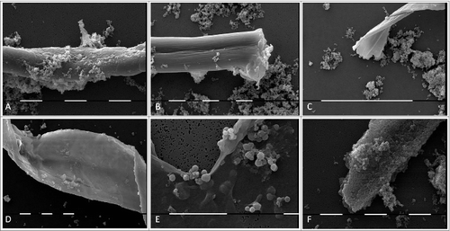
Figure 4. SEM of L-form cells from blood isolate No1 grown in semisolid agar during the second phase/week of cultivation. (A) Cluster of granular forms; (B, C, F) groups of polymorphic granular, oval, rod and elongated filamentous forms; (D, E) typical coccoid and large spherical L-form bodies of different size. Bars: 10 μm (A, B,C, D, E); 1 μm (F).

Figure 5. TEM of L-form cells from blood isolate No1 grown in semisolid agar: (A-C) cell wall deficient cells observed during the first phase/week of cultivation; (D-F) heterogeneous population of reverting bacteria with partially or fully recovered cell walls found during the second phase/week of cultivation.
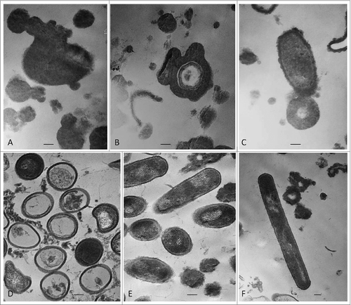
Figure 5. TEM of L-form cells from blood isolate No1 grown in semisolid agar: (A-C) cell wall deficient cells observed during the first phase/week of cultivation; (D-F) heterogeneous population of reverting bacteria with partially or fully recovered cell walls found during the second phase/week of cultivation.

Figure 6. 16S rRNA PCR of blood L-form cultures (L) grown during the second phase/week of cultivation in semisolid agar. Legend: 1, DNA ladder 100 bp; 2,Water; 3, L1; 4, L6; 5, L14 6, L47; 7, L58; 8, L60; 9, L62; 10, L64; 11, L65; 12, L70; 13, L96; 14, L16; 15, L19; 16, L20; 17, L23; 18, L25; 19, L26; 20, L34; 21, L37; 22, L41; 23, L52; 24, L53; 25, L91; 26, L93; 27, M. tuberculosis H37Rv 10ˆ-5; 28, M. tuberculosis H37Rv 10ˆ-3; 29, DNA ladder 100 bp.
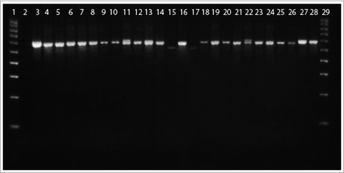
Figure 6. 16S rRNA PCR of blood L-form cultures (L) grown during the second phase/week of cultivation in semisolid agar. Legend: 1, DNA ladder 100 bp; 2,Water; 3, L1; 4, L6; 5, L14 6, L47; 7, L58; 8, L60; 9, L62; 10, L64; 11, L65; 12, L70; 13, L96; 14, L16; 15, L19; 16, L20; 17, L23; 18, L25; 19, L26; 20, L34; 21, L37; 22, L41; 23, L52; 24, L53; 25, L91; 26, L93; 27, M. tuberculosis H37Rv 10ˆ-5; 28, M. tuberculosis H37Rv 10ˆ-3; 29, DNA ladder 100 bp.

Table 1. IS6110 Real Time PCR of blood L-form cultures grown during the second phase/week of cultivation in semisolid agar.

Table 2. Spoligotyping profiles of blood L-form cultures grown during the second phase/week of cultivation in semisolid agar.

Reprints and Corporate Permissions

Please note: Selecting permissions does not provide access to the full text of the article, please see our help page How do I view content?

To request a reprint or corporate permissions for this article, please click on the relevant link below:

Academic Permissions

Please note: Selecting permissions does not provide access to the full text of the article, please see our help page How do I view content?

Obtain permissions instantly via Rightslink by clicking on the button below:

If you are unable to obtain permissions via Rightslink, please complete and submit this Permissions form. For more information, please visit our Permissions help page.