1,119
Views
23
CrossRef citations to date
0
Altmetric
Research Papers

Randomized phase II trial of docetaxel with or without PSA-TRICOM vaccine in patients with castrate-resistant metastatic prostate cancer: A trial of the ECOG-ACRIN cancer research group (E1809)

, , , , , & show all
Pages 2469-2474 | Received 16 Apr 2015, Accepted 09 Jun 2015, Published online: 16 Sep 2015

Figures & data

Figure 1. Study schema.

Figure 1. Study schema.

Table 1. Patient demographics and disease characteristics at study entry

Table 2. Adverse events. Shown are adverse events that were deemed treatment-related, and grade 3 or higher in severity, for the 8 subjects who received assigned treatment

Figure 2. Serum PSA response from patient treated on Arm A. Shown are the serum PSA values (ng/mL) from a patient treated on Arm A with respect to the treatments with the PSA-TRICOM vaccine and docetaxel.
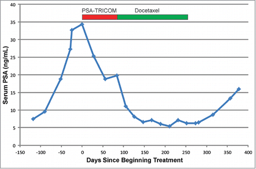
Figure 2. Serum PSA response from patient treated on Arm A. Shown are the serum PSA values (ng/mL) from a patient treated on Arm A with respect to the treatments with the PSA-TRICOM vaccine and docetaxel.

Figure 3. T-cell response evaluation by IFNγ ELISPOT. Cyropreserved PBMC obtained from treated subjects were assessed for IFNγ release following culture with PSA (test antigen, left panel) or phytohemagglutinin (PHA, positive control, right panel) at the individual time points for each subject. Shown are the spot-forming units (sfu) per million PBMC for the antigen-specific conditions subtracting the sfu from media alone.
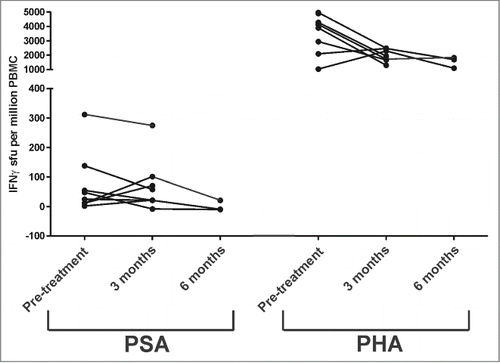
Figure 3. T-cell response evaluation by IFNγ ELISPOT. Cyropreserved PBMC obtained from treated subjects were assessed for IFNγ release following culture with PSA (test antigen, left panel) or phytohemagglutinin (PHA, positive control, right panel) at the individual time points for each subject. Shown are the spot-forming units (sfu) per million PBMC for the antigen-specific conditions subtracting the sfu from media alone.

Figure 4. T-cell response evaluation by intracellular cytokine staining. Cryopreserved PBMC obtained from treated subjects were assessed for cytokine (IFNγ, TNFα, granzyme B (GrB), IL-2, IL-4, IL-10, and IL-17) production by CD4+ (top panels) and CD8+ (bottom panels) T cells following stimulation with PSA (test antigen, left panels) or prostatic acid phosphatase (PAP, control antigen, right panels). Shown is the percentage of CD4+ or CD8+ T cells expressing each cytokine or enzyme at the individual time points for each subject.
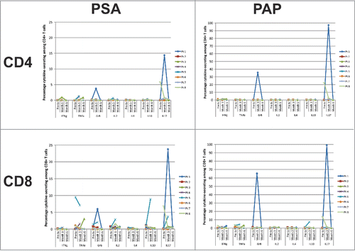
Figure 4. T-cell response evaluation by intracellular cytokine staining. Cryopreserved PBMC obtained from treated subjects were assessed for cytokine (IFNγ, TNFα, granzyme B (GrB), IL-2, IL-4, IL-10, and IL-17) production by CD4+ (top panels) and CD8+ (bottom panels) T cells following stimulation with PSA (test antigen, left panels) or prostatic acid phosphatase (PAP, control antigen, right panels). Shown is the percentage of CD4+ or CD8+ T cells expressing each cytokine or enzyme at the individual time points for each subject.

Reprints and Corporate Permissions

Please note: Selecting permissions does not provide access to the full text of the article, please see our help page How do I view content?

To request a reprint or corporate permissions for this article, please click on the relevant link below:

Academic Permissions

Please note: Selecting permissions does not provide access to the full text of the article, please see our help page How do I view content?

Obtain permissions instantly via Rightslink by clicking on the button below:

If you are unable to obtain permissions via Rightslink, please complete and submit this Permissions form. For more information, please visit our Permissions help page.