Figures & data
Figure 1. The expression of EgG1Y162 in BCG. M: Protein Marker; lanes 1,2: the expression of BCG and rBCG-EgG1Y162 were induced at room temperature for 1 h, respectively; lanes 3, 4: the expression of rBCG-EgG1Y162 were induced for 3 h, 6 h at room temperature, respectively; lanes 5 and 6: the high expression of rBCG-EgG1Y162 could be induced for 3 h at 45°C.
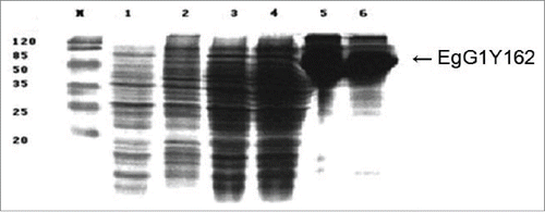
Figure 2. Detected the antigenicity of rBCG-EgG1Y162 by Western blots. M: Protein Marker, lanes 1 to 16: 1–6 serums from rabbits infected by Eg, 7–11 from patients infected by Eg and 12–16 from 5 normal serum samples as negative controls. All of serum samples were diluted 200-fold with PBS.
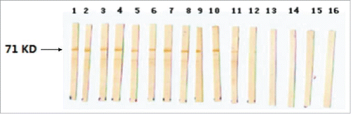
Figure 3. The proliferation of mouse splencytes cultured for 24 h, 48 h, and 72 h in vitro. Microscope images of mouse splencytes stimulated by normal saline, BCG and rBCG-EgG1Y162 for 48 h in vitro. A: normal saline B: BCG C: rBCG-EgG1Y162(I). Compared to normal saline, splenocytes stimulated by rBCG-EgG1Y162 proliferated significantly, while splenocytes stimulated by BCG proliferate slightly. The mean stimulation index for the immunized mice had higher than the index of the negative control mice (P < 0.05). The splenocytes from both groups showed equal responses after stimulated with ConA, suggested that the splencytes were functional and responsive to nonspecific mitogens.
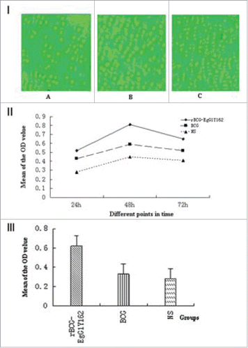
Figure 4. The level of different cytokine in culture supernatants from splenocytes stimulated by rBCG-EgG1Y162 and BCG, respectively. There were no significant differences in TNF-α among each group. Levelsof IL-4, IFN-γ, IL-10, and IL-2 increased significantly in culture supernatants from splenocytes stimulated by rBCG-EgG1Y162. (* means significant difference, P < 0.05).
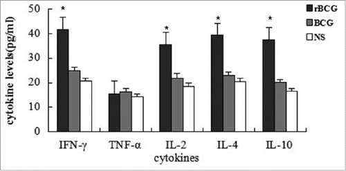
Table 1. The concentrations of different cytokines in culture supernatants from splenocytes stimulated by rBCG-EgG1Y162 and BCG, respectively (μg/ml)
Figure 5. Concentration of the IgG in different groups. IgG sub-classes and the IgE antibodies response against rBCG-EgG1Y162 were detected by ELISA. Serum samples come from BALB/c mice 6 weeks after first immunization. Antibody levels were significant differences between the immunized group and the control group (*P < 0.05, **P < 0.01). Mice immunized by rBCG-EgG1Y162 generated specific high levels of IgG1, IgG2b, and IgG2a.
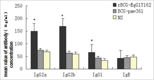
Table 2. The concentration (μg/ml) of the mouse serum antibodies in Balb/C mice sixth weeks after vaccination
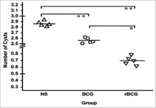
Figure 6. Immune protection of rBCG-EgG1Y162 vaccine against Eg in vivo. Each mouse was immunized with the rBCG-EgG1Y162 or BCG once every 2 weeks, a total of 3 times, respectively. Then challenged with 3000 viable protoscoleces intraperitoneally after the last immunization. The controls groups were immunized with normal saline. The average number of visible cysts decreased in the experimental group (0.7 per mouse), compared to the number in the BCG and normal saline group (control group) (2.86 per mouse).Values are the mean number of cysts ± SEM from 10 mice. *P < 0.05, **P < 0.01 compared to the mean value of each group.