Figures & data
Figure 1. Graphical Schematic of Skin DC Migration to the Cutaneous Lymph Nodes. Epidermal (Langerhans Cells) and dermal skin DC colonize their respective layers of the skin. In both the inflamed and steady state setting, LC and dermal DC enter the lymphatic vessels, before migrating in a CCR7 dependent manner to the local cutaneous draining lymph node bearing either pathogen derived or self antigens. Upon arrival at the lymph node, skin migratory DC enter the paracortical area and present skin derived antigens to T cells. Additionally, post migration, migratory DC can capture and present antigens that have drained freely, such as DEC205 targeted vaccinations. Images were provided by Matthew Woodruff (Wikimedia commons).
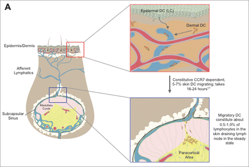
Table 1. Approaches to study skin DC
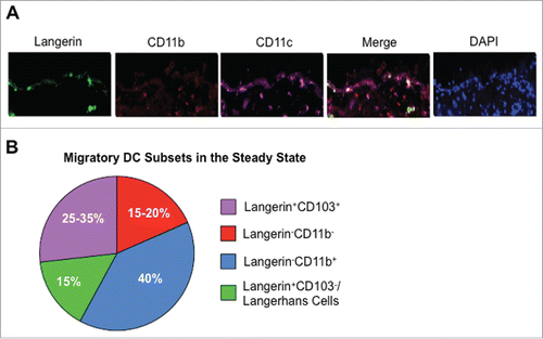
Figure 2. Migratory DC in the Skin and Cutaneous Draining Lymph Node. (A) Murine skin was stained for CD11c (purple), Langerin (green), CD11b (red), and DAPI (blue) in the steady-state. Images were taken at 20 × and are courtesy of Sze-Wah Tse. (B) Proportions of individual migratory DC populations among the total migratory DC subset in the cutaneous draining lymph node in the steady state. Data are adapted from Henri et al.Citation106 and Mollah et al.Citation39