Figures & data
Figure 1. Schematic structures of HA protein of H5N1 and construction and characterization of recombinant HA1-FdFc, HA1-Fc, HA1-Fd, and HA1-His proteins. (A) Schematic structures of HA protein of A/Anhui/1/2005 (H5N1 HA). The HA protein consists of signal peptide (SP, residues 1-12), HA1 (residues 13-325) and HA2 (residues 326-554) fragments. (B) Construction of HA1-FdFc, HA1-Fc, HA1-Fd, and HA1-His protein fragments. The four fragments were constructed by fusing HA1 fragment with Fd and Fc (HA1-FdFc), Fc (HA1-Fc), Fd (HA1-Fd), or His6, respectively. IL2ss, a signal peptide, induces expressed proteins to culture supernatants. The purified protein samples were detected by SDS-PAGE, following by staining with Coomassie Blue (C), or Western blot using HA1-specific HA-7 mAb (D).
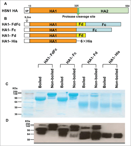
Figure 2. Mouse vaccination procedure and sample collection. Groups of mice were i.n. immunized with HA1-FdFc, HA1-Fc, HA1-Fd, and HA1-His protein, respectively, or PBS, in the presence of Poly (I:C) or CpG adjuvant, or without adjuvant. Mice were immunized 3 times at 3-week intervals. Ten days later, after the last vaccination, mouse sera and lung wash were collected to detect IgG, IgA, and neutralizing antibodies.
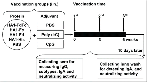
Figure 3. Detection of IgG antibody responses by ELISA in mice immunized with HA1 fusion proteins plus Poly(I:C) or CpG adjuvant. PBS with or without adjuvants was used as the negative control. ELISA plates were coated with HA1-FdFc, HA1-Fc, HA1-Fd, or HA1-His protein, respectively (A), or full-length HA1-His protein (B), and IgG antibody was detected using mouse sera (1:3,200) from 10 days post-last vaccination. The data are presented as A450 ± SD of 4 mice per group. The *, ** and *** indicate the significant difference with P < 0.05, 0.01 and 0.001, respectively, between the groups with or without adjuvants.
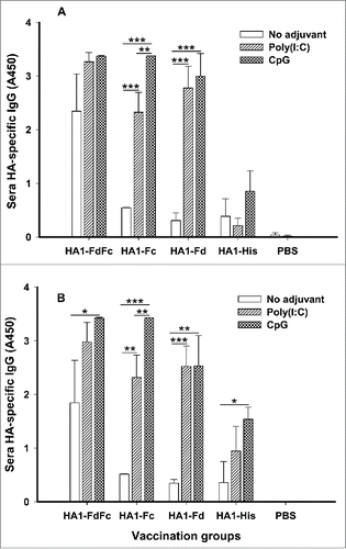
Figure 4. Detection of IgG1 and IgG2a subtype antibody responses by ELISA in mice immunized with HA1 fusion proteins plus Poly(I:C) or CpG adjuvant. PBS with or without adjuvants was used as the negative control. The ability of IgG1 (A) and IgG2a (B) antibodies to bind H5N1 HA1 protein was detected using sera from 10 days post-last vaccination. The data are presented as A450 ± SD of 4 mice per group. The *, ** and *** indicate the significant difference with P < 0.05, 0.01 and 0.001, respectively, between the groups with or without adjuvants.
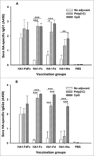
Figure 5. Detection of IgA antibody responses by ELISA in mice immunized with HA1 fusion proteins plus Poly(I:C) or CpG adjuvant. PBS with or without adjuvants was used as the negative control. The ability of IgA to bind H5N1 HA1 protein was detected using mouse lung wash (A) and sera (B) from 10 days post-last vaccination. The data are presented as A450 ± SD of 4 mice per group. The *, ** and *** indicate the significant difference with P < 0.05, 0.01 and 0.001, respectively, between the groups with or without adjuvants.
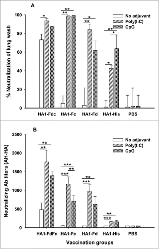
Figure 6. Comparison of neutralizing antibodies in lung wash (A) and sera (B) of mice immunized with HA1 fusion proteins plus Poly(I:C) or CpG adjuvant. PBS with or without adjuvants was used as the negative control. The lung wash (1:1,000) and sera from 10 days post-last immunization were pooled in each group and tested for neutralizing antibodies. The data of panel A are presented as mean percentages of inhibition of duplicated wells. The data are presented as mean ± SD of duplicated wells. The *, ** and *** indicate the significant difference with P < 0.05, 0.01 and 0.001, respectively.