Figures & data
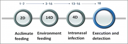
Figure 1. Pathological changes detected by HE staining. (A) Uninfected mice in a normal environment. (B) Infected mice in a normal environment. (C) Uninfected mice in the wet environment. (D) Infected mice in the wet environment. (E) Uninfected mice in the warm-wet environment. (F) Infected mice in the warm-wet environment.
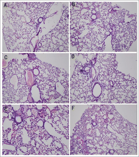
Figure 2. Serum cytokine levels detected by ELISA. (A) Serum levels of IL-4 in each group. (B) Serum levels of IFN-γ in each group. (C) Serum levels of IL-10 in each group. (D) Serum levels of IL-17 in each group. The cytokine levels were elevated during infection, and the levels in the group B animals were higher than group A. In the wet and wet-hot environments, the cytokines levels were suppressed, and the levels of groups D and F were lower than those of group B. There was no significant difference between group D and group F. *P < 0.05; ns, not significant.
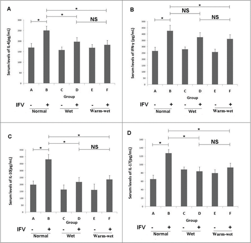
Figure 3. The T cell subset proportions were detected by FCM. (A) Th1/Th2 proportions. In the normal environment, the proportion of group B was higher than that of group A, the proportions of Th1/Th2 in groups D and F were lower than group B. (B) Th17/Treg proportions. In the normal environment, the proportion of group B was higher than that of group A, the proportions of Th17/Treg cells in groups D and F were lower than group B. There were no significant differences between group D and group F. *P < 0.05; ns, not significant.
Figure 4. Expression of the RLH signal pathway as detected by RT-PCR and Western blotting. (A) Relative expression of IRF3 mRNA. (B) Relative expression of IRF7 mRNA. (C) Relative expression of RIG-1 mRNA. (D) Relative expression of IPS-1 mRNA. (E) Relative expression of TBK-1 mRNA. (F) Expression IPS-1 and RIG-1 proteins. The expression of the RLH pathway in groups D and F were lower than group B. The relative expression of mRNA in the uninfected animals in the normal environment was set as 1. *P < 0.05; ns, not significant.
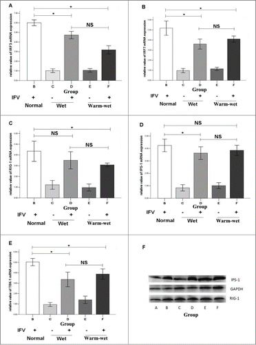
Table 1. Experimental groups of this study. All the groups were feeding in the manual climatic box and half of the rats were infected by virus
Figure 5. The timeline of model establish. For the first 2 days, the mice were kept in our P2 laboratory to acclimate to the feeding environment. On the third day, mice were introduced to the 3 different environments. On the thirteenth day, the mice were infected with the FM1 virus through nasal inhalation for 4 days; on the 18 day, the mice were executed for viral detection.