
Figures & data
Figure 1. Electron micrographs of the highly purified NoV GII.4 VLPs (A), GI.3 VLPs (B) and recombinant RV VP6 protein (C) examined by FEI Tecnai F12 electron microscope (Philips Electron Optics, Holland) following negative staining with 3% uranyl acetate (pH 4.6).
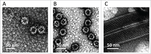
Figure 2. NoV GII.4 (A, B) and GI.3 (C, D) genotype-specific serum IgG antibody responses induced with 0.3 μg or 3 μg of GII.4 or GI.3 VLPs alone or with 0.3 μg doses in a combination with 10 μg of RV VP6. Control (Ctrl) mice received carrier (PBS) only. OD490nm values of GII.4- (A) and GI.3-specific (C) antibodies in 1:100 diluted sera of individual mice are shown with the horizontal line representing the mean OD490nm value of the experimental group. GII.4- (B) and GI.3-specific (D) end-point titers of the groups of mice expressed as the geometric mean titers of the reciprocal of the highest sample dilution giving a positive reading. Error bars represent 95% confidence intervals, CIs. Groups were compared by Mann-Whitney U-test or Fisher's exact test and p values determined.
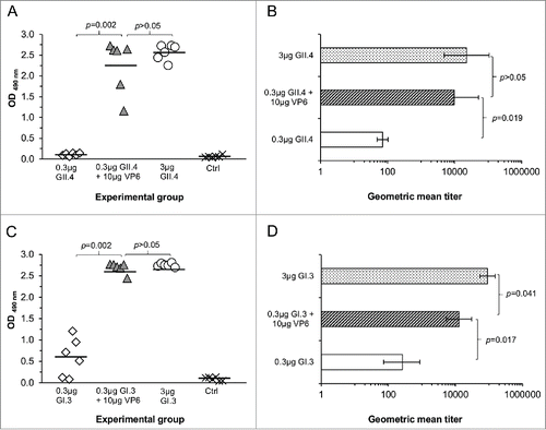
Table 1. Experimental and control groups of immunized mice. Mice were immunized intramuscularly (IM) at day 0 and 21 with the indicated dose and terminated at day 35.
Figure 3. Cross-reactivity of serum IgG antibodies to NoV GII.4 NO (A), GII.4 Sydney (B) and GI.1 (C) in mice immunized with 0.3 μg of GII.4 or GI.3 VLPs alone or in a combination with 10 μg of RV VP6. Control (Ctrl) mice received carrier only (PBS). Mean ODs of the experimetal groups with standard errors are shown.
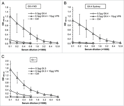
Figure 4. RV VP6-specific IgG antibody responses in sera of mice immunized with 0.3 μg of GII.4 (A) or GI.3 (B) VLPs in a combination with 10 μg VP6. End-point titration curves of each immunized mice and mean curve of the control (Ctrl) mice are shown.
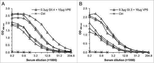
Figure 5. Blocking of NoV GII.4 VLP (A and B) and GI.3 VLP (C and D) binding to human type A saliva (A and C) and synthetic HBGAs (B and D) by the sera of mice immunized with 0.3 μg or 3 μg of GII.4 or GI.3 VLPs alone or with 0.3 μg doses in a combination with 10 μg of RV VP6. Synthetic HBGA H type 1 was employed for GII.4 (B) and Lea HBGA for GI.3 VLP (D) binding. Serum from mice receiving PBS was used as a negative control (Ctrl). Results are shown as the mean % blocking with standard errors of 2 – 3 independent experiments. A dashed line indicates 50% cut-off.Citation47
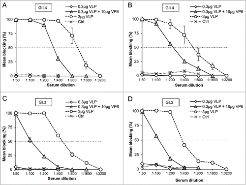
Figure 6. RV VP6-specific IL-4 and IFN-γ production by T-cells. RV Wa cell culture antigen and recombinant VP6 protein were used to stimulate IL-4 ((A)and B) and IFN-γ (C) production from the splenocytes of mice immunized with 0.3 μg of GII.4 or GI.3 VLPs alone or in a combination with 10 μg of RV VP6. Control (Ctrl) mice received carrier only (PBS). Results are expressed as the mean spot forming cells (SFC)/106 cells of the duplicate wells with standard errors. The experiments were repeated 2 or more times with similar results. A dashed line indicates the cut-off limit >50 SFC/106 splenocytes.
