Figures & data
Figure 1. Schematic diagram of the chimeric human/mouse MHC class I gene. (A) Schematic diagram of the monochain chimeric human/mouse MHC class I gene showing the HHD structure of the chimeric human α1, α2, and β2m HLA-A11/murine H-2 α3 molecule. The murine H2 α3 domain is covalently linked to human β2m via a 15 amino acid linker. (B) Liner representation of the final construct.
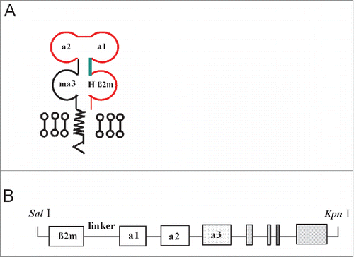
Figure 2. Genotype of HLA-A11/DR1 Tg mice and flow cytometric analysis of transgenic HLA molecules expressed on the surface of mouse immune cells. Genomic DNA purified from HLA-A11/DR1 Tg mice was analyzed by PCR using primers specific for HLA-A11, HLA-DR1, mouse β2m, and H-2 IAβ. (A) Identification of HLA-A11 (upper panel) and HLA-DR1 (lower panel). Tg mice, HLA-A11/DR1 Tg mice; NC, Negative control; PC, Positive control; Blank, dH2O. (B) Identification of mouse β2m(upper panel) and H-2 IAβ (lower panel).Tg mice,HLA-A11/DR1 Tg mice; NC, Negative control; PC, Positive control; F1, Heterozygote control; Blank, dH2O.(C and D) Splenocytes from HLA-A11/DR1 (red histogram), H-2-I/II knockout mice (black histogram), and wild-type C57BL/6 (blue histogram) mice were isolated and stained with a PE-anti-human-HLA-ABC antibody (C) or an FITC-anti-human-HLA-DR antibody (D) to detect HLA-A11 and HLA-DR1 expression, respectively.
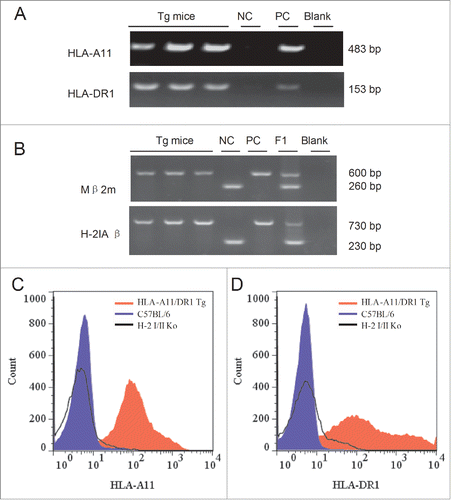
Figure 3. Flow cytometric analysis of peripheral CD8+ and CD4+ (T)lymphocytes. (A-D) Splenocytes from HLA-A11/DR1 mice (A), HLA-A11 mice (B), HLA-DR1 mice (C), and wild-type C57BL/6 mice (D) were isolated. CD3+ T lymphocytes were gated by staining with an FITC-anti-CD3 mAb and CD8+ T lymphocytes were gated by staining with a PEcy5-anti-CD8 mAb. (E-H) Splenocytes from HLA-A11/DR1 mice (E), HLA-A11 mice (F), HLA-DR1 mice (G), and wild-type C57BL/6 mice (H) were isolated. CD3+ T lymphocytes were gated by staining with a PE-anti-CD3 mAb, and CD4+ T lymphocytes were gated by staining with an APC-anti-CD4 mAb.
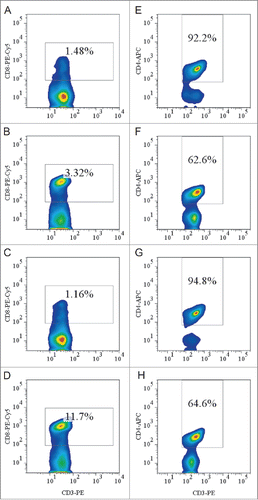
Figure 4. HBs-specific antibody- and cell-mediated responses after immunization with a recombinant HBsAg vaccine. (A) Sera were collected from immunized HLA-A11/DR1 Tg mice and wild-type C57BL/6 mice, and the titers of anti-HBs (IgG) antibodies were compared with those of PBS-immunized (hollow bar) mice in an ELISA test. (B) HBs epitope-specific IFN-γ production by cytotoxic T lymphocytes was examined by measuring the response of both HLA-A11/DR1 Tg mice and wild-type C57BL/6 mice to a recombinant HBs vaccine or PBS.
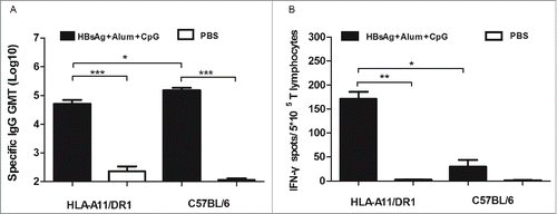
Figure 5. HIV-specific antibody and CD8+ (T)cell responses after immunization with a recombinant HIV-1 protein. (A) HIV-specific antibody titers in HLA-A11/DR1 Tg and C57BL/6 mice immunized with a recombinant HIV-1 protein or PBS. (B) HIV-specific IFN-γ production by cytotoxic T lymphocytes was examined by measuring responses to the recombinant HIV-1 protein in immunized mice (PBS-immunized mice were used as control).
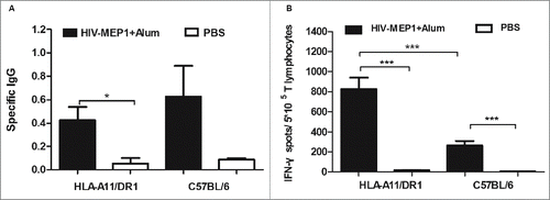
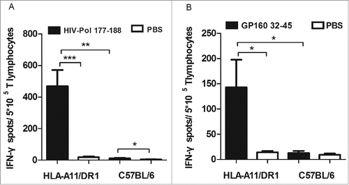
Figure 6. HLA-A11 restricted cytotoxic responses in HLA-A11 transgenic mice. Responses to (A) the HLA-A11-restricted epitope, HIV-Pol177–188, and (B) the HLA-A11-restricted epitope, HIV-GP32–45, were assessed in immunized HLA-A11 transgenic and wild-type C57BL/6 mice by counting the number of IFN-γ-secreting spots in ELISPOT assays.