Figures & data
Table 1. Levels of antigen-specific IgA antibody responses at systemic and mucosal sites.
Table 2. Levels of antigen-specific IgG antibody responses at systemic and mucosal sites.
Table 3. Levels of antigen-specific IgM antibody responses at systemic and mucosal sites.
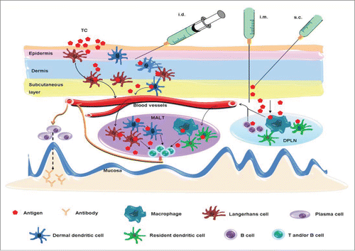
Figure 1. Potential mechanisms of systemic vaccination-induced mucosal antibody responses. Intradermal (i.d.) or transcutaneous (TC) immunization activates Langerhans cells and dermal dendritic cells in the epidermis and dermis of skin, which then migrate to the mucosa-associated lymphoid tissue (MALT) where they present the antigen to CD4+ T cells and B cells. An antigen delivered by i.m. or s.c. route mainly diffuses to the draining peripheral lymph nodes (DPLN) where it activates APCs, such as B cells, dendritic cells and macrophages. Mucosal antibody responses are triggered when they reach to the MALT and present the antigen to CD4+ T cells and B cells. A free antigen may migrate to MALT directly.