1,195
Views
12
CrossRef citations to date
0
Altmetric
Review

Plasmodium falciparum gametocyte transit through the cutaneous microvasculature: A new target for malaria transmission blocking vaccines?

Pages 3189-3195 | Received 03 Feb 2016, Accepted 21 Apr 2016, Published online: 19 Jul 2016

Figures & data

Figure 1. The five morphologically distinct stages (I – V) of P. falciparum gametocytes in giemsa stained, methanol fixed blood films. Early subpellicular cytoskeleton formation in stage II results in D-shaped parasites that enlarge in stage III to slightly distend the erythrocyte. Dramatic elongation and pointed ends characterize stage IV gametocytes that ultimately mature into stage V gametocytes. Characteristic morphology of this final stage includes an elongated parasite with rounded ends and a length to width ratio of ∼3:1. Dark pigmented hemozoin crystals are present in multiple gametocyte developmental stages, and are generated from the biocrystallization of hematin, a toxic intermediate derived from the parasite digestion of hemoglobin.

Figure 1. The five morphologically distinct stages (I – V) of P. falciparum gametocytes in giemsa stained, methanol fixed blood films. Early subpellicular cytoskeleton formation in stage II results in D-shaped parasites that enlarge in stage III to slightly distend the erythrocyte. Dramatic elongation and pointed ends characterize stage IV gametocytes that ultimately mature into stage V gametocytes. Characteristic morphology of this final stage includes an elongated parasite with rounded ends and a length to width ratio of ∼3:1. Dark pigmented hemozoin crystals are present in multiple gametocyte developmental stages, and are generated from the biocrystallization of hematin, a toxic intermediate derived from the parasite digestion of hemoglobin.
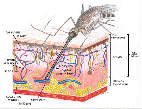
Figure 2. Illustration of the architecture of the cutaneous microcirculation and the fascicle of a biting anopheline mosquito. The vessel diameters of the arterioles, collecting venules, terminal arterioles, and capillary loops are denoted along with the fascicle length and width.

Figure 2. Illustration of the architecture of the cutaneous microcirculation and the fascicle of a biting anopheline mosquito. The vessel diameters of the arterioles, collecting venules, terminal arterioles, and capillary loops are denoted along with the fascicle length and width.

Reprints and Corporate Permissions

Please note: Selecting permissions does not provide access to the full text of the article, please see our help page How do I view content?

To request a reprint or corporate permissions for this article, please click on the relevant link below:

Academic Permissions

Please note: Selecting permissions does not provide access to the full text of the article, please see our help page How do I view content?

Obtain permissions instantly via Rightslink by clicking on the button below:

If you are unable to obtain permissions via Rightslink, please complete and submit this Permissions form. For more information, please visit our Permissions help page.