Figures & data
Figure 1. Modification altered expression levels of IE1 and IE2. Western blot analysis of HEK293 cells transfected with DNA plasmids expressing wild-type IE1 or modified IE1 (A); wild-type IE2, IE2 with two alanine substitutions (IE2(H2A)), modified IE2 (mIE2), or modified IE2 with alanine substitutions (mIE2(H2A)) (B).
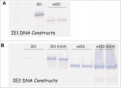
Table 1. Summary of CMV antigen constructs.
Figure 2. Expression of wild-type versus modified HCMV antigens by Ad6 vectors. Western blot analysis of Per.C6 cell lysates from Ad6-Mock, Ad6-pp65 and Ad6-mpp65 (A: lane 1, 2 and 3, respectively); Ad6-IE1 and Ad6-mIE1 (B: lane 1 and 2, respectively); Ad6-IE2 and Ad6-mIE2 (C: lane 1 and 2, respectively).
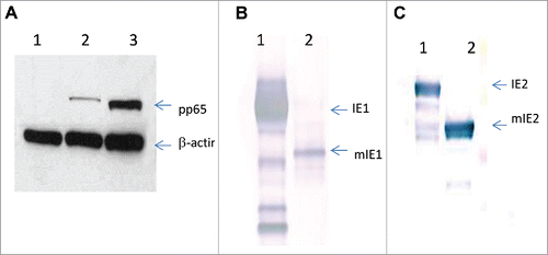
Figure 3. Subcellular localization of wild-type versus modified HCMV antigens by indirect immunofluorescent staining. MRC-5 cells infected with Ad6-pp65 (A), Ad6-mpp65 (B), Ad6-IE1 (C), Ad6-mIE1 (D), Ad6-IE2 (E) and Ad6-IE2 (F) were stained with antigen-specific antibodies and antibody specific to Sp100. Cells were also nucleus stained with DAPI. Pictures were acquired for individual staining and overlaying using confocal microscopy as described in Materials and methods.
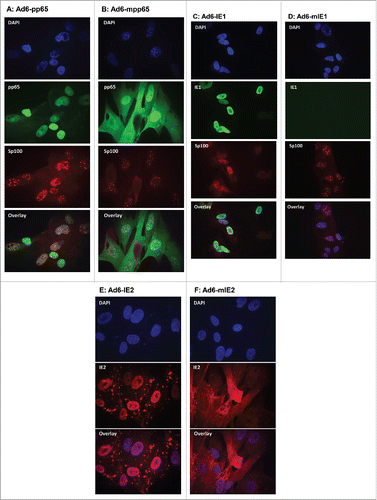
Figure 4. Comparable immunogenicity of wild-type versus modified HCMV antigens in mice. C57BL/6 × Balb/c F1 mice (n = 10) were immunized with the indicated vaccine and dose, and the spleen cells from four mice were pooled and assessed for IFN-γ secretion in response to re-stimulation with corresponding antigen pp65 (A), IE1 (B) and IE2(C) peptide pools in ELISPOT assays. Open circles in plots represent vaccines with wild-type antigens and open triangle represent vaccines with modified antigens. DMSO at the same concentration as in peptide pools was used as negative control (dashed lines). Number of spot forming cells (SFC) on y axis was plotted against vaccine doses of viral particles (vp) on x axis.
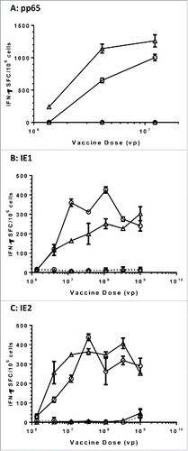
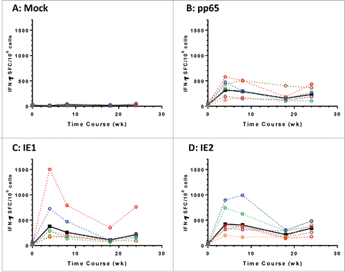
Figure 5. Modified HCMV antigens immunogenic in nonhuman primates. T cell responses in six rhesus macaques immunized with a mixture of Ad6 vector vaccines at day 0 and wk 20. The mixture contains Ad6-mpp65, Ad6-mIE1 and Ad6-mIE2 at 1 × 10¹⁰ vp per construct. PBMCs were assessed for IFN-γ secretion pre-vaccination and at weeks 4, 8, 18 and 24 post vaccination in ELISPOT assay. PBMCs were stimulated with DMSO control (A), pp65 peptide pool (B), IE1 peptide pool (C) or IE2 peptide pool (D). IFN-γ secreting cell numbers per 1 × 10⁶ PBMCs for each monkey were plotted in colored dashed lines and geometric mean values from all monkeys were plotted in solid black line. Number of spot forming cells (SFC) on y axis was plotted against time points of PBMC sampling.