Figures & data
Figure 1. RSV ΔF-specific systemic humoral immune responses in mice. (A) Serum IgG1 and IgG2a titers before challenge (B/C), (B) Serum IgG1 and IgG2a titers after challenge (A/C) with RSV, (C) Serum VN antibody titers determined before (B/C) and after (A/C) RSV challenge, and (D) Numbers of IFN-γ and IL-5 secreting splenocytes determined in response to in vitro restimulation with ΔF protein. BALB/c mice were immunized once IN with ΔF formulated with TriAdj and challenged 3 weeks later with RSV. Control groups were immunized with PBS and challenged with RSV (Placebo) or mock-challenged (Placebo/mock). ELISA titers are expressed as the reciprocal of the highest dilution resulting in a value of two standard deviations above the negative control serum. Virus neutralization titers are expressed as the highest dilution of serum that resulted in <50% of cells displaying cytopathic effects. Cytokine secreting cell numbers are expressed as the difference in the number of spots between ΔF-stimulated wells and medium-control wells. Bars indicate median values with interquartile ranges. **P < 0.01; ***P < 0.001.
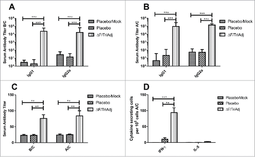
Figure 2. Mucosal immune responses to RSV ΔF protein in mice after challenge with RSV. The IgA titers (A), virus titers (B), percentages of ΔF-specific CD8+ T cells (C), and ΔF-specific IFN-γ secreting CD8+ T cells (D) were measured in the lung after RSV challenge. Mice were immunized and challenged as described in the legend for . ELISA titers are expressed as the reciprocal of the highest dilution resulting in a value of two standard deviations above the negative control serum. Virus replication in the lungs is expressed as pfu per gram of lung tissue. The proportion of RSV F-specific CD8+ T cells was determined in the lung by KYKNAVTEL-MHC I pentamer staining. IFN-γ expression by CD8+ T cells was determined by intracellular cytokine staining. Bars represent median values with interquartile ranges. *P < 0.05; **P < 0.01; ***P < 0.001.
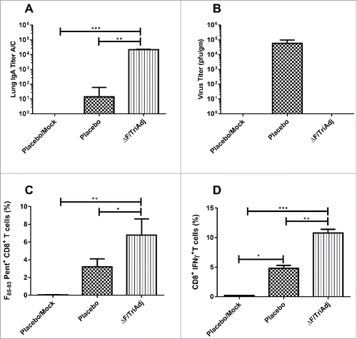
Figure 3. Long-term systemic immune responses to RSV ΔF protein. IgG (A) and VN (B) titers were measured at different times after vaccination. BALB/c mice were immunized once IN with ΔF formulated with TriAdj and a control group was immunized with PBS (Placebo). ELISA titers are expressed as the reciprocal of the highest dilution resulting in a value of two standard deviations above the negative control serum. Virus neutralization titers are expressed as the highest dilution of serum that resulted in <50% of cells displaying cytopathic effects. Symbols represent median values with interquartile ranges.
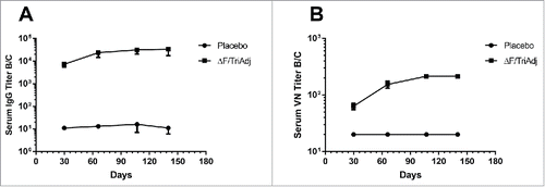
Figure 4. Systemic immune responses to RSV ΔF protein. (A) Serum IgG1 and IgG2a titers before challenge (B/C), (B) Serum IgG1 and IgG2a titers after RSV challenge (A/C), (C) Serum VN antibody titers after RSV challenge (A/C), and (D) Numbers of IFN-γ and IL-5 secreting splenocytes determined in response to in vitro restimulation with ΔF protein. BALB/c mice were immunized once IN with ΔF formulated with TriAdj and challenged with RSV on day 150. Control groups were immunized with PBS and challenged with RSV (Placebo) or mock-challenged (Placebo/mock). ELISA titers are expressed as the reciprocal of the highest dilution resulting in a value of two standard deviations above the negative control serum. Virus neutralization titers are expressed as the highest dilution of serum that resulted in <50% of cells displaying cytopathic effects. Cytokine secreting cell numbers are expressed as the difference in the number of spots between ΔF-stimulated wells and medium-control wells. Bars indicate median values with interquartile ranges. ***P < 0.001.
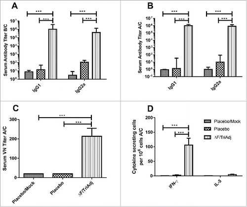
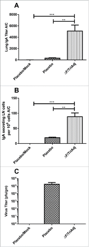
Figure 5. Long-term mucosal immune responses to RSV ΔF protein. IgA titers (A), numbers of IgA secreting LN cells (B) and virus titers (C) were determined after RSV challenge (A/C). Mice were immunized and challenged with RSV as described in the legend for . ELISA titers are expressed as the reciprocal of the highest dilution resulting in a value of two standard deviations above the negative control serum. IgA secreting cell numbers are expressed as the difference in the number of spots between ΔF-stimulated wells and medium-control wells. Virus replication in the lungs is expressed as pfu per gram of lung tissue. Bars represent median values with interquartile ranges. **P < 0.01; ***P < 0.001.