652
Views
1
CrossRef citations to date
0
Altmetric
Research Paper

Intradermal delivery of a fractional dose of influenza H7N9 split vaccine elicits protective immunity in mice and rats

, , , , , , , , , & show all
Pages 623-629 | Received 28 Aug 2017, Accepted 26 Dec 2017, Published online: 12 Feb 2018

Figures & data

Figure 1. The ventral sites of rats were wiped and injected ID with 100 µL of 10% ink-PBS using the MP-0.1 system. The animals were euthanized, and their skin was dissected and lifted to confirm the accuracy of the injection (A, B). On day 0, the rats were immunized IM (syringe) or ID (MP-0.1 system) with 3, 9, or 15 µg of HA, or PBS, in a total volume of 100 µL, and body weight changes were recorded daily (C).
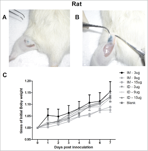
Figure 1. The ventral sites of rats were wiped and injected ID with 100 µL of 10% ink-PBS using the MP-0.1 system. The animals were euthanized, and their skin was dissected and lifted to confirm the accuracy of the injection (A, B). On day 0, the rats were immunized IM (syringe) or ID (MP-0.1 system) with 3, 9, or 15 µg of HA, or PBS, in a total volume of 100 µL, and body weight changes were recorded daily (C).

Figure 2. Six-to-eight-week-old BALB/c mice (n = 6) and SD rats (n = 6) were twice immunized ID or IM with 3, 9, or 15 µg of HA of the split H7N9 influenza vaccine at 2-week intervals, and serum samples were collected at 2, 3, and 4 weeks post-boosting for HI antibody measurements (A, B). Mice and rats were immunized with the H7N9 influenza vaccine at 0 and 14 days, and serum HI antibody titers and IgG subtypes were assayed at 2 and 4 weeks post-boosting (C).
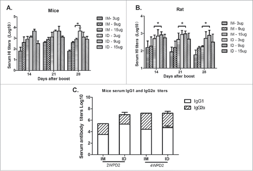
Figure 2. Six-to-eight-week-old BALB/c mice (n = 6) and SD rats (n = 6) were twice immunized ID or IM with 3, 9, or 15 µg of HA of the split H7N9 influenza vaccine at 2-week intervals, and serum samples were collected at 2, 3, and 4 weeks post-boosting for HI antibody measurements (A, B). Mice and rats were immunized with the H7N9 influenza vaccine at 0 and 14 days, and serum HI antibody titers and IgG subtypes were assayed at 2 and 4 weeks post-boosting (C).

Figure 3. BALB/c mice (n = 6) were immunized IM or ID with 3 or 15 µg HA of the H7N9 influenza vaccine, and serum HI antibody titers were measured beginning at day 0 (A). Thirty rats were injected with 3 µg of HA of the H7N9 influenza vaccine, or saline as a control, using a syringe or the MP-0.1 system, and skin biopsies were obtained at 0, 1, 2, 3, and 7 days for assessment of inflammatory cell infiltration into the dermis and muscular layer (B).
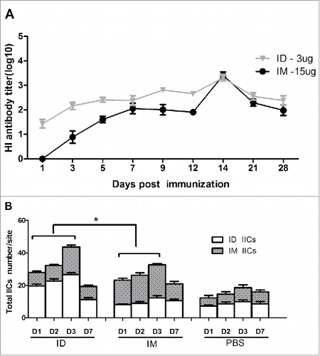
Figure 3. BALB/c mice (n = 6) were immunized IM or ID with 3 or 15 µg HA of the H7N9 influenza vaccine, and serum HI antibody titers were measured beginning at day 0 (A). Thirty rats were injected with 3 µg of HA of the H7N9 influenza vaccine, or saline as a control, using a syringe or the MP-0.1 system, and skin biopsies were obtained at 0, 1, 2, 3, and 7 days for assessment of inflammatory cell infiltration into the dermis and muscular layer (B).

Figure 4. The H7N9 split vaccine was labeled with Cy5 for visualization of antigen deposition and metabolism in vivo. Rats were immunized ID using MP-0.1 or IM using a syringe and anesthetized with 4% isoflurane. Fluorescence signals were visualized at 1 hour, 1 day, 2 days, 5 days, and 7 days, and background auto-fluorescence was subtracted (A). Fluorescence intensity over time (B). At 1, 24, and 48 h, three rats per group were euthanized, and the kidney, liver, spleen, and thymus were removed and subjected to fluorescence imaging (C, D, E, F).
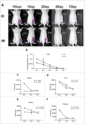
Figure 4. The H7N9 split vaccine was labeled with Cy5 for visualization of antigen deposition and metabolism in vivo. Rats were immunized ID using MP-0.1 or IM using a syringe and anesthetized with 4% isoflurane. Fluorescence signals were visualized at 1 hour, 1 day, 2 days, 5 days, and 7 days, and background auto-fluorescence was subtracted (A). Fluorescence intensity over time (B). At 1, 24, and 48 h, three rats per group were euthanized, and the kidney, liver, spleen, and thymus were removed and subjected to fluorescence imaging (C, D, E, F).

Figure 5. At 0 and 14 days, BALB/c mice were immunized IM with PBS or 15 or 3 µg HA, or they were immunized ID with 3 µg HA of the influenza H7N9 split vaccine. Two weeks later, they were challenged IN with a lethal dose of wild-type A/AnHui/1/2013 H7N9 virus (5.0 × 10³ TCID50 pfu). Body weight was monitored daily (n = 6), and pathological changes (n = 3) in the lungs were analyzed at 7 days after challenge (A, B, C, D, E).
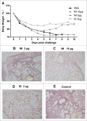
Figure 5. At 0 and 14 days, BALB/c mice were immunized IM with PBS or 15 or 3 µg HA, or they were immunized ID with 3 µg HA of the influenza H7N9 split vaccine. Two weeks later, they were challenged IN with a lethal dose of wild-type A/AnHui/1/2013 H7N9 virus (5.0 × 10³ TCID50 pfu). Body weight was monitored daily (n = 6), and pathological changes (n = 3) in the lungs were analyzed at 7 days after challenge (A, B, C, D, E).
Supplemental material

KHVI_A_1423156_Supplemental.zip

Download Zip (7.6 MB)

Reprints and Corporate Permissions

Please note: Selecting permissions does not provide access to the full text of the article, please see our help page How do I view content?

To request a reprint or corporate permissions for this article, please click on the relevant link below:

Academic Permissions

Please note: Selecting permissions does not provide access to the full text of the article, please see our help page How do I view content?

Obtain permissions instantly via Rightslink by clicking on the button below:

If you are unable to obtain permissions via Rightslink, please complete and submit this Permissions form. For more information, please visit our Permissions help page.