Figures & data
Figure 1. EE significantly improved the activity of AD mice in the open field. In the 60 minutes open field test, the results were analyzed as a session every 10 minutes. The results showed that EE significantly enhanced the ambulation of AD mice during the whole experiment(A). EE significantly increased the time of central lattice movement (B), rears (C) and fine (D) of AD mice. *P < 0.05, compared with wild type C57; #P < 0.05, compared with AD mice.
Figure 2. EE significantly improved the learning and memory ability in AD mice. In the 3rd and 4th day, the acquisition of place navigation, wild type C57 and AD mice reared in EE used significantly less time to find the platform. In the 7th day, the reversal of place navigation, wild type C57 and AD mice reared in EE performed better (A). In the 5th day, the spatial probe, the AD mice reared in the EE spent significantly more times to through the target platform than the AD group (B), meanwhile the percentage of swimming time and distance in the target quadrant were significantly higher than the AD group (C, D). There was no difference in swimming speed between the three groups. *P < 0.05, compared with wild type C57; #P < 0.05, compared with AD mice.
Figure 3. EE significantly reduced the Aβ plaques and peptides in the brain of AD mice. Immunofluorescence showed that, compared with AD mice, most of the larger plaques in hippocampus (A, B) and cortex (D, E) area decrease in EE mice, while the deposition of some dispersion have also disappeared. Analysis of quantitative image showed the Aβ deposition in the hippocampus (C) and cortex (F) of AD mice in EE were significantly. The soluble/insoluble Aβ1–40/Aβ1–42 peptides (G, H) show EE reduced the levels of Aβ peptides in AD mice. *P < 0.05, **P < 0.01, compared with AD mice.
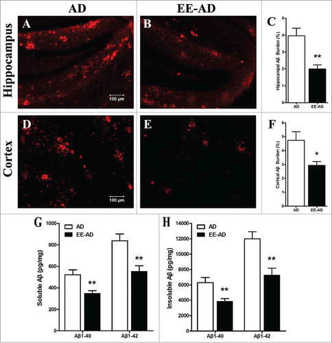
Figure 4. Changes of the content of Ach, ChAT and AchE in the hippocampus (A, B, C) and cortex (D, E, F) of the mice. Compared with the control group, the Ach and ChAT in the brain of AD mice decreased significantly, while the content of AchE increased significantly. The AD mice fed in EE showed that these changes were relieved in varying degrees, but there wash no statistical difference compared with the AD mice. *P < 0.05, **P < 0.01, ***P < 0.001, compared with wild type C57.
Figure 5. Detection of inflammatory and neurotrophic cytokines level in mice. The results showed that the level of inflammatory factors IL-1β (A), IL-6 (B), TNF-α (C) and IFN-γ (D) in serum of AD mice were significantly higher than those of wild type C57 mice, while trophic factor BDNF (E), NGF (F) and IGF-1 (G) in hippocampus were lower. By reareing in EE, the inflammatory factors decreased while the trophic cytokines increased significantly. *P < 0.05, **P < 0.01, compared with wild type C57; #P < 0.05, compared with AD mice.
Figure 6. The mice previously raised in EE represent no difference with the AD mice, In the place navigation (A), the wild type C57 spent significantly less time to find the platform. In the spatial probe (B-D), the wild type C57 spent more times and distance in the target quadrant than the other two groups. *P < 0.05, compared with wild type C57.
Figure 7. The inflammatory and neurotrophic cytokines in mice. The level of inflammatory factors IL-1β (A), IL-6 (B), TNF-α (C) and IFN-γ (D) in serum of AD mice which were previously reared in EE were higher than the controls, and the trophic factors BDNF (E), NGF (F) and IGF-1 (G) in hippocampus were lower. *P < 0.05, **P < 0.01, ***P < 0.001, compared with wild type C57.