Figures & data
Figure 1. PTg but not PTc induces cord blood T cell response. Cord blood mononuclear cells (n = 7) were stimulated with PT antigens or TLR agonists (TLR3 agonist, Poly I:C; TLR4 agonist, MPLA and TLR7/8 agonist, R848)as indicated. Surface expression of CD4 T cell early activation marker CD69 (a), intracellular IL17 (b) and IL2 (c) levels and, surface co-expression of OX40 and CD25 (d) were determined by flow cytometry. Results are expressed as mean ± standard deviation. * p < 0.05, **p < 0.01, ***p < 0.001, ****p < 0.0001 denotes significance compared to unstimulated control.
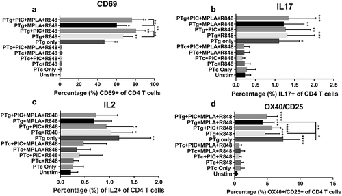
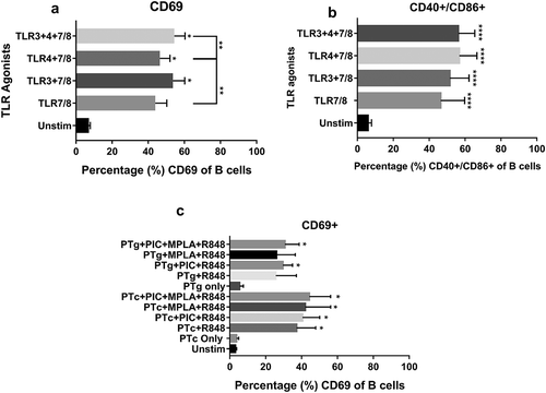
Figure 2. TLR agonist combinations but not PT induces cord blood B cell activation. Cord blood mononuclear cells (n = 4) were stimulated with PT antigens or TLR agonists (TLR3 agonist, Poly I:C; TLR4 agonist, MPLA and TLR7/8 agonist, R848) as indicated. Flow cytometric analysis of B cell – CD69 (a) as well as costimulatory marker CD40/CD86 expression (b) were measured after TLR agonist stimulation. B cell – CD69 expression in the presence of PT antigens (c) were also tested. Results are expressed as mean ± standard deviation. * p < 0.05, **p < 0.01, ***p < 0.001, ****p < 0.0001 denotes significance compared to unstimulated control.