Figures & data
Table 1. Adverse effects of nonsurgical therapy.
Figure 1. Patient with osteoradionecrosis of the mandible from radiation therapy.
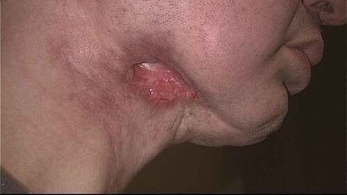
Figure 2. Patient with T1N2A oropharyngeal cancer status post definitive chemoradiation. (a) Distal chip video laryngoscopy revealed post-treatment radiation edema with no evidence of mucosal disease delineated by red arrows. (b) NBI revealed a mucosal abnormality delineated by blue arrows. Biopsy was consistent with high-grade dysplasia. Image is borrowed with permission from Dr Peter Belafsky.
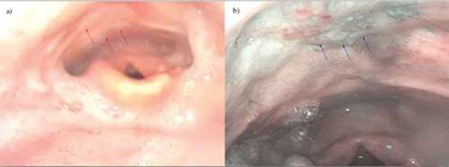
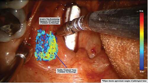
Figure 3. This intraoperative image shows the use of lifetime tissue autofluorescence for identification of pathologic mucosa. A laser is aimed at the surface and the signal returning from the tissue is interpreted. The signal for pathologic mucosa has been shown to be distinctly different than normal and is demonstrated by a different shade on the color scale. Such an approach could be very useful in cancer screening. However, small submucosal lesions may avoid detection.