Figures & data
Figure 1. HBV virus amplification & characterization from HepG2.2.15 cells.
(a) Western blot for detection of M-HBsAg in cell lysate of HepG2.2.15 cells. Detection of M-HBsAg in the cell lysate of HepG2.2.15 cells. 10, 20, 30 and 40 μg of cell lysate was loaded in the lanes (b) Detection of S-HBsAg. ELISA for detection of s-HBsAg in the supernatant and cell lysate of HepG2.2.15 cells (c) Immunofluorescence detection of HBsAg preS2 antigen in HepG2.2.15 cells. (d) Quantification of HBV-DNA copies in the supernatant of HepG2.2.15 cells by qPCR. Cell culture supernatant from HepG2.2.15 cells was harvested and concentrated 100 fold with amicon centrifugation. DNA from 50 ul concentrated supernatant was extracted and HBV DNA copies in the supernatant were quantified using synthetic HBV DNA as standard. DNA extracted from Vero cells was used as a negative control. (e) Agarose gel electrophoresis of qPCR reaction system to determine presence of 81 bp amplicon.
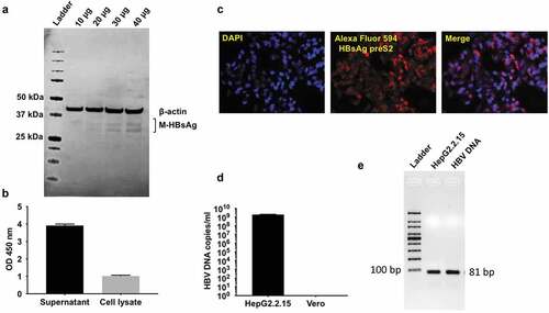
Figure 2. HBV-DMAb expression in vitro and in vivo.
(a) Cloning of HBsAg DMAb in pVax1 expression vector. (b) 293 T cells were transfected with HBV-DMAb or pVax1. Human IgG expression in culture supernatant and cell lysate was quantified by ELISA (n = 3 biological replicates, mean ± SD). (c) Western blot analysis of human IgG heavy and light chain in reduced supernatant and cell lysates of 293 T cells transfected with HBV-DMAb or pVax1. HBV-DMAb purified from serum from mice immunized with HBV-DMAb was loaded as a control (right). (d) HBV-DMAb expression in CAnN.Cg-Foxn1 nu/Crl nude mice after electroporation-enhanced intramuscular inoculation with 100 μg HBV-DMAb. Sera were collected up to 35 days post administration (n = 5 mice per group, mean ± SD). (e) Mean expression level in mice sera of HBV-DMAb. (f) ELISA binding of mouse serum collected 21 days post administration of HBV-DMAb to plasma purified native HBsAg.
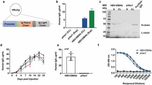
Figure 3. HBV-DMAb binds a specific epitope of HBsAg.
(a) IFA to detect HBV-DMAb binding to HBV in HepG2.2.15 cells. Cells were incubated with serum from mice immunized with HBV-DMAb or pVax1 followed by staining with the Alexa fluor 594 antibody conjugate. The cell nuclei were stained with DAPI. (b) Western blot of HBsAG after SDS-PAGE separation under reducing (denatured) conditions. No binding of HBV-DMAb was observed when reduced HBsAg was used in SDS-PAGE.(c) Native PAGE and western blot analysis of HBsAg using sera from mice immunized with HBV DMAb. Detection of DMAb binding to purified HBsAg was performed using goat anti-human IgG-HRP conjugate by chemiluminescence. No binding of HBV-DMAb was observed when denatured HBsAg was used in SDS-PAGE. However, the DMAb strongly bound to native HBsAg in Native-PAGE which indicates that antibody binds to a conformational epitope of HBsAg.
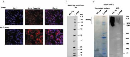
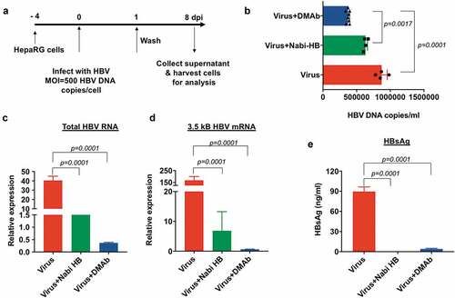
Figure 4. HBV-DMAb neutralizes HBV and blocks infection of HepaRG cells.
(a) Schematic of infection of HepaRG cells with HBV. (b) Quantification of viral load in the culture supernatant of HepaRG cells by qPCR. (c) Relative quantification of total HBV RNA and (d) 3.5 kb pregenomic RNA in HepaRG cells by qRT-PCR. Values represent mRNA expression relative to cells treated with Nabi-HB. (e) Measurement of HBsAg secreted in the culture medium by ELISA. Quantity of antibody used for neutralization assay: Nabi-HB: 1.25 mg and HBV-DMAb: 10 µg.