
Figures & data
Figure 1. In vitro characterizations of UV-inactivated DENV2 TMC NPs
Diameter and zeta potential of UV-inactivated DENV2 loaded TMC NPs were determined by zetasizer (a). Cellular uptake of UV-inactivated DENV2 TMC NPs (3 or 15 μg of encapsidated UV-inactivated DENV2) or sUV-inactivated DENV2 (15 μg) by MoDCs at 24 and 48 h post-treatment was evaluated by intracellular staining with mouse anti-DENV2 E protein antibody (3H5). Stained cells were then detected by flow cytometry; (b) dot plot analysis, (c) percentage of DENV-2 positive cells and (d) mean fluorescent intensity (MFI). (e) The uptaken UV-inactivated DENV2 of treated cells after 48 h post-treatment was imaged by a fluorescent microscope (200× and 400× magnifications). Data are represented as mean ± SD (n = 3). * indicates a significant difference in sUV-inactivated DENV2 and UV-inactivated DENV2 TMC NPs at indicated time points (p < .05). sUV-DENV2 and UV-DENV2 TMC NPs represent a sUV-inactivated DENV2 and an UV-inactivated DENV2 TMC NPs, respectively .
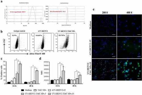
Figure 2. TMC NPs assist UV-inactivated DENV2 in enhancing the maturation of MoDCs
Cells were stimulated with TMC NPs (20 or 100 μg) or UV-inactivated DENV2 TMC NPs (20 or 100 μg of TMC containing 3 or 15 μg of UV-inactivated DENV2 antigen) or sUV-inactivated DENV2 (15 μg) for 48 h. After incubation, treated cells were harvested and stained with antibodies specific to activating markers of MoDCs. The percentage of positive cells and expression of CD80 (a and e), CD83 (b and f), CD86 (c and g) and HLA-DR (d and h) were measured by flow cytometry. Data are shown as mean ± SD (n = 3). *, # indicate significant differences of MoDCs exposed to UV-inactivated DENV2 TMC NPs and TMC NPs at 20 or 100 μg of TMC, respectively, and “a” indicates a significant difference between UV-inactivated DENV2 TMC NPs and sUV-inactivated DENV2 (p < .05). sUV-DENV2 and UV-DENV2 TMC NPs represent a sUV-inactivated DENV2 and an UV-inactivated DENV2 TMC NPs, respectively .
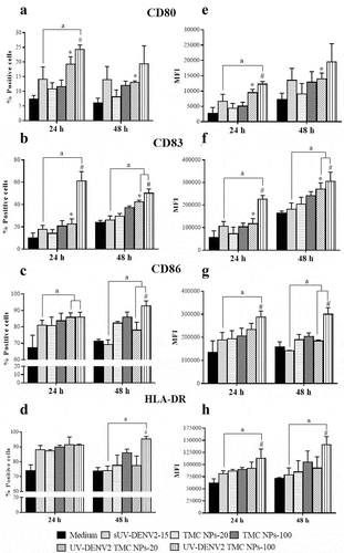
Figure 3. Cytokine and chemokine profiles of UV-inactivated DENV2 TMC NPs treated MoDCs
Cells were treated with TMC NPs (20 or 100 μg) or UV-inactivated DENV2 TMC NPs (20 or 100 μg of TMC containing 3 or 15 μg of encapsidated UV-inactivated DENV2) or sUV-inactivated DENV2 (15 μg) for 48 h. At indicated time points, culture supernatants of treated cells from three different donors were harvested and subjected for secreted cytokine/chemokine quantification using Bio-Plex based assay. (a) Inflammatory cytokines, (b) chemokines, (c) growth factors, (d) Th-1 related cytokines, (e) Th-2 related cytokines and (f) type I interferon. Results are presented as mean ± SD. *, # indicate significant differences of MoDCs treated with UV-inactivated DENV2 TMC NPs and TMC NPs at 20 or 100 μg of TMC, respectively (p < .05). sUV-DENV2 and UV-DENV2 TMC NPs represent a sUV-inactivated DENV2 and an UV-inactivated DENV2 TMC NPs, respectively .
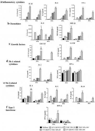
Figure 4. Immunization with UV-inactivated DENV2 TMC NPs up-regulates antibody production
(a) Mice were vaccinated via i.p. injection with three doses (15 days apart) of sUV-inactivated DENV2 or UV-inactivated DENV2 TMC NPs (10 or 20 μg/dose). (b) DENV-2 specific IgG titers in mouse sera by days 0, 14, 29, 37 and 45 were quantitated by indirect ELISA. (c) Sera diluted at 1:2000 on day 45 post-immunization were subjected to detection of DENV-2 specific IgG using capture ELISA. Serum IgG1 (d) and IgG2a (e) were measured by indirect ELISA and results are presented as OD values. Data are shown as mean ± SEM (n = 5). *, # indicate significant differences between sUV-inactivated DENV2 and UV-inactivated DENV2 TMC NPs at an equal dose of antigen (p < .05). sUV-DENV2 and UV-DENV2 TMC NPs represent a sUV-inactivated DENV2 and an UV-inactivated DENV2 TMC NPs, respectively .
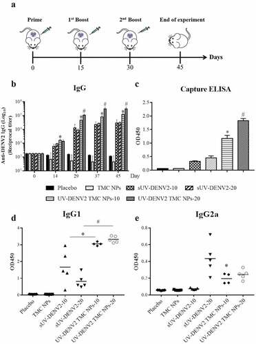
Figure 5. Production of neutralizing antibody against DENV-2 upon UV-inactivated DENV2 TMC NPs or sUV-inactivated DENV2 immunization
Sera from mice were collected on days 14, 29, 37 and 45. The titers of NAb (Neut50) against DENV-2 reference strain (16681) were quantified by PRNT. Data are shown as mean ± SEM (n = 5). *, # indicate significant differences between sUV-inactivated DENV2 and UV-inactivated DENV2 TMC NPs at an equal dose of antigen (p < .05). sUV-DENV2 and UV-DENV2 TMC NPs represent a sUV-inactivated DENV2 and an UV-inactivated DENV2 TMC NPs, respectively .
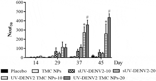
Figure 6. Neutralizing enhancement of DENV-2 immune serum upon treatment with complement
(a) serum from each immunized mice harvested on day 45 was subjected to PRNT in the presence or absence of rabbit complement (1:200 dilution). Data are represented as mean of Neut50 ± SEM (n = 5). (b) DENV-2 infected BHK-21 cells were incubated with the ten-fold dilutions of mouse sera before addition of complement. The supernatant was collected for the detection of released lactate dehydrogenase (LDH). Data are mean ± SD (n = 3). “a” indicates a significant difference between the addition of complement and non-complement control (p < .05). *, # indicate significant differences between sUV-inactivated DENV2 and UV-inactivated DENV2 TMC NPs at the same doses of antigen (p < .05). sUV-DENV2 and UV-DENV2 TMC NPs represent a sUV-inactivated DENV2 and an UV-inactivated DENV2 TMC NPs, respectively .
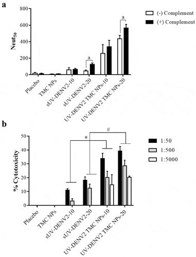
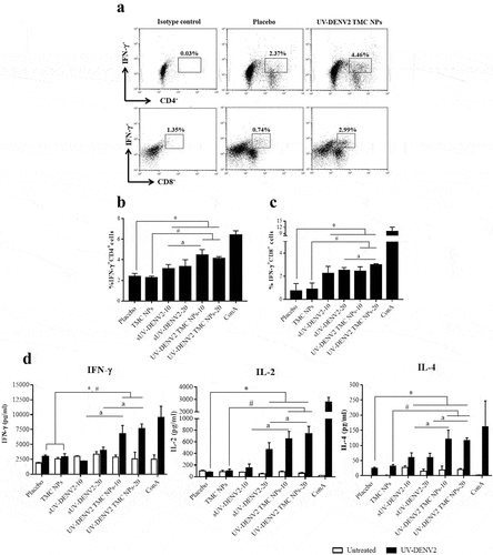
Figure 7. Cell-mediated immune responses elicited by UV-inactivated DENV2 TMC NPs or sUV-inactivated DENV2 vaccination
Splenocytes isolated from mice on day 45 post-vaccination were stimulated with 10 μg/ml of UV-inactivated DENV2 antigen for 72 h. Stimulated cells were harvested and double-stained with antibodies corresponding to CD4 or CD8 and IFN-γ, and followed by analyzing with flow cytometry. The levels of cytokines in supernatant from splenocyte cultures were determined by ELISA. Results are shown in (a) dot plot analysis, (b) frequency of IFN-γ⁺CD4⁺, (c) IFN-γ⁺ CD8⁺ cells and (d) cytokine production including IFN-γ (72 h), IL-2 (24 h) and IL-4 (48 h). Data are mean ± SEM (n = 3). *, # indicate significant differences in groups of mice vaccinated with UV-inactivated DENV2 TMC NPs compared to placebo or TMC NPs, respectively. “a” indicates a significant difference in a group of mice vaccinated with sUV-inactivated DENV2 and UV-inactivated DENV2 TMC NPs at same doses (p < .05). sUV-DENV2 and UV-DENV2 TMC NPs represent a sUV-inactivated DENV2 and an UV-inactivated DENV2 TMC NPs, respectively .
Supplemental material