Figures & data
Table 1. Measurements of immunoglobulin concentration in lavages of various mucosal secretions. Though IgA comprises the majority of antibody at most surfaces, substantial concentrations of IgG are found at many mucosal interfaces, and predominates in cervicovaginal secretions. The concentrations have been rounded to two significant figures
Table 2. Measurements of normalized diffusion coefficients for Abs in cervical mucus. If a particle diffuses in mucus as fast as it diffuses in buffer, Dmuc/Dpbs = 1. Abs with multiple entries are results of independent experiments. IgM, sIgA aggregates, and IgM Fc complex with removed Fab domains are slowed moderately in mucus. IgA and IgG were hardly slowed in mucus
Figure 1. Schematic of Ab-mediated trapping. Virus alone can freely diffuse through the mucin fibers. Weak interactions between the Fc portion of the Ab and the mucin allow individual Ab to diffuse freely. As the Fab arms bind to antigen and Ab accumulates on the viral surface, Ab-virus complexes become tightly complexed to the matrix.
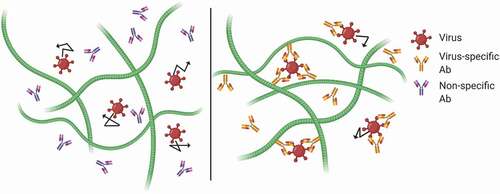
Figure 2. Deglycosylation of IgG abolishes trapping of HSV in CVM. (a) Fc removal from anti-HSV1, confirmed by SDS-PAGE. (b) Deglycosylation of anti-HSV1, confirmed by lectin-ELISA. (c) Effective diffusivity of HSV1 in the CVM treated with intact, Fc-removed, and deglycosylated IgG. Reproduced with permission from.Citation21 Copyright 2014 Springer nature.
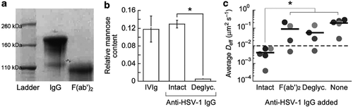
Figure 3. Transient vs. stable Ab-matrix bonds. Diffusion of 200 nm polyethylene glycol (PEG)-conjugated latex nanoparticles in biotinylated Matrigel® modified with neutravidin. (a) Representative traces of nanoparticles in Matrigel® with no added IgG (control), anti-PEG IgG (IgG), or biotinylated anti-PEG IgG (IgG-biotin) exhibiting effective diffusivities within one SEM of the ensemble average at a timescale of 1 s. (b) Distributions of the mean logarithms of individual particle effective diffusivities (Deff) at a timescale of 0.2667 s. Log (Deff) values to the left of the dashed line correspond to particles with displacements of less than 100 nm (i.e., roughly the particle diameter) within 0.2667 s. (c) Ensemble-averaged geometric mean square displacements (<MSD>) as a function of timescale, (d) mean Deff of all particles in each condition, and (e) fraction of mobile nanoparticles in Matrigel® treated with different IgG. Reproduced under creative commons license from.Citation44
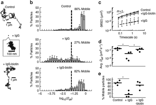
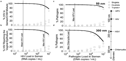
Figure 4. Simulations of virion collisions at different viral loads and particle sizes. The fraction of virions over the first 12 hours post ejaculation as a function of virion density in semen which have undergone no collision (singlets), one collision (doublets) or two or more collisions (triplets+) for all HIV virions (a) in semen/CVM mixture and (b) that have reached the vaginal epithelium. (c–d) The fraction pf (c) 50 nm pathogens and (d) 300 nm pathogens in diameter that have diffused across CVM and reached the vaginal epithelium. Solid line represents an exponential function approximation of the virion load arriving at the epithelial layer that experiences no collisions with other virions. Modified and reproduced under creative commons license from.Citation68