Figures & data
Figure 1. (a–b) Wide-field fundus photographs showed radial retinal wrinkles (white arrows) surrounding the optic disc and fovea OU at presentation (C-D), which disappeared one month after presentation.
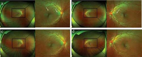
Figure 2. (a–b) OCT also showed radial retinal wrinkles (white arrows) at presentation, (c–d) which then disappeared one month after presentation. (e–f) FA revealed optic disc swelling (white arrows) and macular edema OU at presentation.
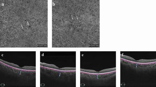
Figure 3. (a–b) OCTA showed diffuse ischemia per se (white arrows) on the choriocapillaris OU at presentation. (c–d) The choroidal thickness of the subfovea increased to 519 μm OD and 608 μm OS at presentation and (e–f) then decreased to 352 μm OD and 365 μm OS three months later.