Figures & data
Figure 1. The eukaryotic expression vector pCAG-scFv4F5-GFP was constructed and then transfected into A549 cells.
Figure 2. The expressed intracellular antibody can inhibit H5N1 virus propagation in A549 cells.
Figure 3. Full-length human anti-H5N1 virus IgG1 antibody-expressing vectors were constructed and then stably transfected into CHO-DG44 cells.
Figure 4. The purified full-length human IgG1 antibody has high specificity, sensitivity and affinity against H5N1 virus.
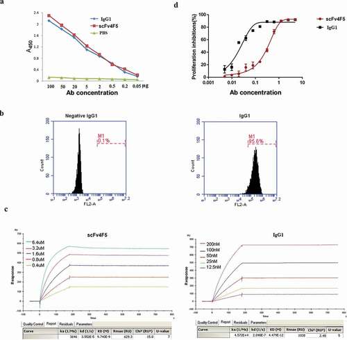
Figure 5. The combination of intracellular and extracellular antibodies has a better protective effect in a mouse model challenged with H5N1 virus.
Figure 6. The secretion of cytokines and the apoptosis-related proteins increased after administration of the antibody combination.