Figures & data
Figure 1. Membrane surface protein of SARS-CoV-2 and the mechanism of SARS-CoV-2 invading cells. A. S1 subunit recognizes and binds ACE2 on host cell membrane. B. Proteases from host cells disassemble ACE2 and activate S2 subunit C. Activated S2 subunit mediates the fusion of virus envelope and host cell membrane, and then the virus invades the cell.
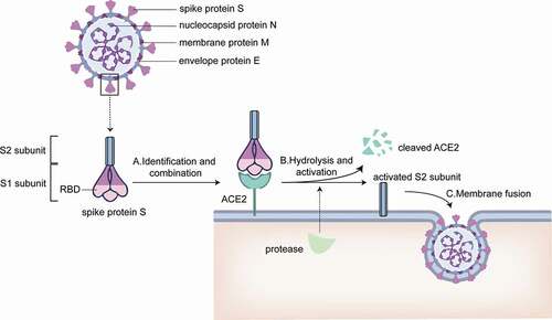
Figure 2. Synthesis of the mRNA vaccine. A. mRNA of target protein is extracted from SARS-CoV-2. B. Targeted mRNA is transcribed into double-helix DNA through reverse transcription; C. DNA strands combine with plasmid. D. Plasmid incorporating fragments of DNA are amplified in Escherichia coli; E. the target DNA is sheared from many cloned plasmids and purified; F. the double stranded DNA (dsDNA) template is unwound by enzymes and transcribe into mRNA; G. the final assembly of the vaccine is achieved by enclosing mRNA with liposomes.
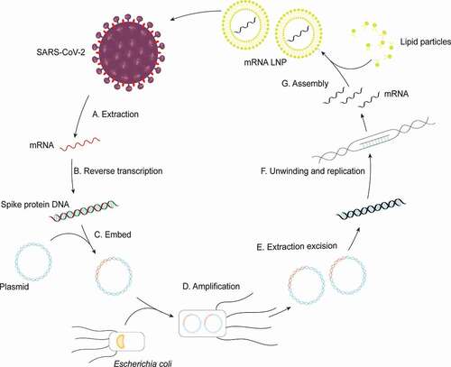
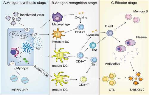
Figure 3. Mechanism of inactivated vaccine and mRNA vaccine. A. Antigen synthesis stage: Myocytes capture inactivated virus from inactivated vaccine and target mRNA LNP from the mRNA vaccine; B. Antigen recognition stage: (1) Antigen presenting cell including DC cells and macrophages recognize the antigen phagocytosed from muscle cells and present it to DC cells; (2) Common features: Both vaccines activate CD4+T cell immunity through the MHC class II pathway; (3) mRNA vaccine advantages: mRNA LNP can activate more CD8+T cells because of the presence of the MHC class I pathway; C. Effector stage: CTL and B cells are activated and destroy the virus. a. Ribosome b. endoplasmic reticulum c. golgiapparatus d. lysosome.