Figures & data
Figure 1. MRI T1 sequence of the head and neck region.
Head and neck MRI showed a tumor mass (4 × 3 cm) on the right piriform fossa. After neoadjuvant chemotherapy, there was shrinkage of the tumor mass (3 × 2 cm) and no recurrence after surgery with postoperative concurrent chemoradiotherapy.
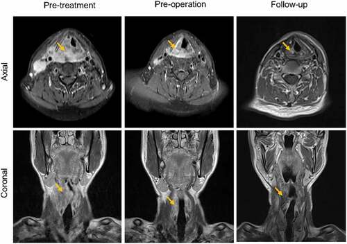
Figure 2. Hematoxylin-Eosin (HE) staining and immunohistochemistry of tumors.
HE staining of the primary tumor and hepatic tumor; immunohistochemistry of the primary tumor for programmed death-ligand 1 (PD-L1) (PD-L1 control and negative control), combined positive score (CPS)≈10; immunohistochemistry of the hepatic tumor for PD-L1 (PD-L1 control and negative control), CPS ≈75. Original magnification ×40.
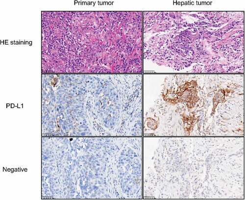
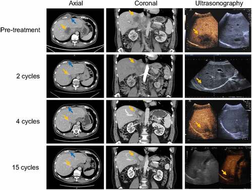
Figure 3. Contrast-enhanced CT and ultrasonography of the abdomen.
Abdominal contrast-enhanced CT and ultrasonography showed hepatic metastasis (5 × 5 cm, orange arrow) and a subcutaneous mass on the right chest wall (blue arrow). After two cycles of immunotherapy, the subcutaneous mass disappeared, and the hepatic metastatic tumor decreased by 46%. After four cycles, the hepatic metastatic tumor reached near complete remission, maintaining 15 cycles of immunotherapy.