Figures & data
Figure 1. Summary of patient treatment history.
As shown in the figure, the red arrow indicated right inguinal lymph node (lesion A). The blue arrow indicated left inguinal lymph node (lesion B), and yellow arrows pointed to locally recurrent lesions (lesion C).
Ia: On February 27, 2020, bilateral inguinal lymph nodes and local recurrent lesions with tumor broken and bleeding. Ib-c: Pelvic CT showed disease progression. IIa: On August 27, 2020, the tumor ruptured and healed gradually, after 2 cycles of immunotherapy combined with chemotherapy.IIb-c: Lesions A and B were assessed as partial response according to RECIST version 1.1, as shown in the pelvic CT image. IIIa: On November 5, 2020, lesion C increased significantly, after 3 cycles of maintenance monotherapy treatment with sintilimab. IIIb-c Lesion A and B were assessed to be stable disease, while lesion C progressed. Ⅳa: On May 6, 2021, the lesion C was significantly reduced after treatment with sintilimab plus gemcitabine. Ⅳb-c: All three lesions were evaluated as partial response, as shown in the pelvic CT image.
Abbreviations: SD: disease progression; PR: partial response; TP: paclitaxel combined with cisplatin chemotherapy regimen
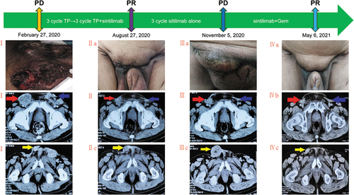
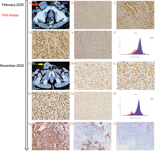
Figure 2. Different lesions represented MMR heterogeneity during treatment.
Ia: Right inguinal lymph node metastasis(Named lesion A) and left inguinal lymph node metastasis (Named lesion B) were indicated by the red and blue arrows, respectively. Ib-Ie: Immunohistochemical detection of mismatch repair protein(MMR) in lesion A showed MLH1 (-), MSH2 (+), MSH6 (+), PMS2 (-),respectively. If: The alteration of BTA-25 site detected by next generation sequencing (NGS)method was confirmed as Microsatellite Instability-Low (MSH-L) status.IIa:Local recurrent lesions (Named lesion C) was indicated by the yellow arrows.IIb-IIe: Immunohistochemical detection of mismatch repair protein (MMR) in lesion C showed MLH1 (+), MSH2 (+), MSH6 (+), PMS2 (+),respectively.IIf: The secondary biopsy specimens detected by NGS were confirmed as Microsatellite stability (MSS) status. IIg: The Lesion C had a PD-L1 tumor proportion score 60% and combined positive score 70 (clone 22C3). IIi-IIh: PD-L1 expression positive control and negative control, respectively.