Figures & data
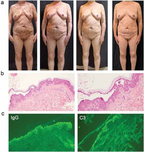
Figure 1. The clinical photographs, Pathological sections, and direct immunofluorescence of patients in Case 1. (a) the clinical photograph taken on admission to the hospital, three weeks after admission, and seven month follow-up (left to right). (b) Histopathological image of the lesions: intraepithelial cleavage with detached keratinocytes primarily localized just under the stratum corneum. (c) Intercellular deposition of immunoglobulin G (IgG) and complement component (C3) by direct immunofluorescence microscopy: spinosum intercellular deposition of complement IgG and C3.
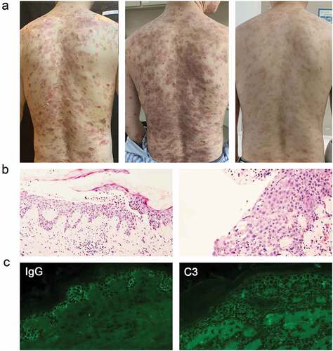
Figure 2. The clinical photographs, Pathological sections, and direct immunofluorescence of patients in Case 2. (a) the clinical photograph taken on admission to the hospital, four weeks after admission, seven weeks after admission, and five month follow-up (left to right). (b) Histopathological image of the lesions: subcorneal split directly below the stratum corneum. (c) Intercellular deposition of immunoglobulin G (IgG) and complement component (C3) by direct immunofluorescence microscopy: spinosum intercellular deposition of complement IgG and C3.