Figures & data
Figure 1. Our decisional matrix for the diagnosis/suspicion of VIPIT, clinical data, i.e. suspicion of thrombosis after AZD 1222 vaccination was the main cause for undergoing this matrix.
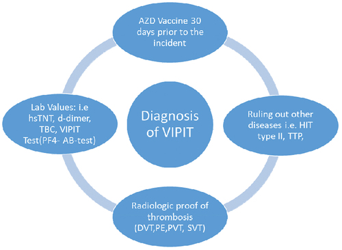
Table 1. Overview over important lab values of the five patients, based on IgG alone (7), measured values based on IgG and IgM.Citation12.
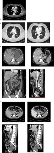
Figure 2. FCT scans of patients with suspected or confirmed VIPIT: The first scan (a) shows the pulmonary embolism in patient 1, the next two scans (b) shows the pulmonary embolism inpatient 3 and its diffuse pattern. The third scan series (c) show the mesenterial thrombosis in patient 4, and the last four scans (d) shows the mesenterial thrombosis and its extent in patient 5, with a positive anti PF4-test. They both show an extensive portal vein thrombosis (red arrow), best seen in (c) when comparing jugular and mesenterial vein contrast and best seen in d, in the sagittal view, when comparing arterial and venous phase. Both patients (c and d) show a extensive diffuse mesenteric thrombosis originating from the complete thrombosis of the splenic vein, indicating a possible immunologic origin with potential thrombotic chain reaction.