
Figures & data

Figure 1. PRISMA flow diagram for study selection.
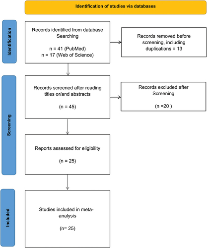
Table 1. Patients from respective case reports included in the analysis.
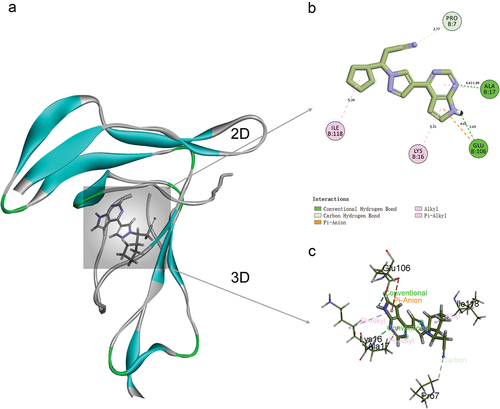
Figure 2. Ruxolitinib was bound to IL-2 Rα by molecular docking. (a) mode of binding of ruxolitinib to IL-2 Rα. (b) two-dimensional interaction between ruxolitinib and IL-2 Rα, circles represent amino acid residues, numbers inside are amino acid abbreviations and numbers, lines represent interactions, colors represent interaction types, and numbers on lines represent distances. (c) 3D structure of ruxolitinib binding to IL-2 Rα.
Supplemental material