
Figures & data
Figure 1. Musculoskeletal ultrasonographic scans (a and b) and MRIs (c and d) of knee after noting a significant foot drop. (a) longitudinal scan view, lobulated feature of deep peroneal nerve (white arrows), (b) two-week follow-up scan view, slightly enlarged in size for two weeks. (HOF: head of fibula) (c) axial T2-weighted image shows about 11 cm extent of longitudinal cystic lesion (asterisk) along common peroneal nerve and articular branch with high signal intensity. T2 high signal intensity changes probably due to denervation are shown on tibialis anterior and extensor hallucis longus muscles (arrow head). (d) sagittal T2-weighted image shows cyst (asterisk) along the track of the common peroneal nerve (white arrows) around the fibular head.
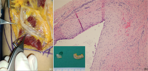
Figure 2. Intraoperative findings and histological confirmation. (a) the common peroneal nerve runs around the fibular head, and a longitudinal and lobulated mass (arrowhead) is observed inside the nerve sheath. (b) Pathological examination reveals a cystic structure containing mucinous or gelatinous fluid and lined by flattened or cuboidal cells, confirming the diagnosis as a ganglion cyst. (hematoxylin and eosin stain, ×100).
Data availability statement
The data supporting the findings of this study are available upon request from the corresponding author.