1,328
Views
5
CrossRef citations to date
0
Altmetric
ADDENDUM

Cloning, soluble expression, and purification of the RNA polymerase II subunit RPB5 from Saccharomyces cerevisiae

, , , &
Pages 62-66 | Received 19 Nov 2014, Accepted 08 Dec 2014, Published online: 21 Jan 2015

Figures & data

Figure 1. PCR amplification and restriction digestion of S. cerevisiae rpb5 gene. Lane 1: DNA ladder. Lane 2: PCR product (648 bp). Lane 3: pSK+-rpb5 digested with Bam HI & Hind III; upper band (3 Kb) is pSK+ vector and lower band (648 bp) is the rpb5 gene. Lane 4: pET28a(+)-rpb5 was restricted with BamHI & Hind III; upper band (5.3 Kb) shows pET28a vector while the lower band (648 bp) shows rpb5 gene. 1 % agarose gel electrophoresis was used to visualize the bands.
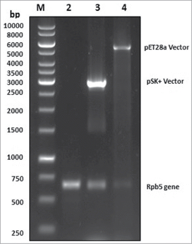
Figure 1. PCR amplification and restriction digestion of S. cerevisiae rpb5 gene. Lane 1: DNA ladder. Lane 2: PCR product (648 bp). Lane 3: pSK+-rpb5 digested with Bam HI & Hind III; upper band (3 Kb) is pSK+ vector and lower band (648 bp) is the rpb5 gene. Lane 4: pET28a(+)-rpb5 was restricted with BamHI & Hind III; upper band (5.3 Kb) shows pET28a vector while the lower band (648 bp) shows rpb5 gene. 1 % agarose gel electrophoresis was used to visualize the bands.

Figure 2. Rpb5 protein expression, solubility and purification analysis. Protein samples were separated by 12 % SDS-PAGE, and stained with CBB. Lane 1: Molecular weight marker. Lane 2: Rpb5 Un-induced control lysate. Lane 3: Rpb5 Induced lysate. Lane 4: Rpb5 protein pellet fraction after lysis. Lane 5: Soluble fraction after lysis. Lane 6: purified Rpb5 protein.
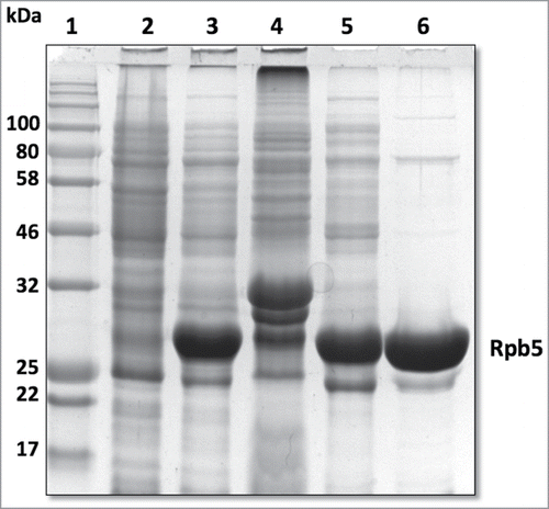
Figure 2. Rpb5 protein expression, solubility and purification analysis. Protein samples were separated by 12 % SDS-PAGE, and stained with CBB. Lane 1: Molecular weight marker. Lane 2: Rpb5 Un-induced control lysate. Lane 3: Rpb5 Induced lysate. Lane 4: Rpb5 protein pellet fraction after lysis. Lane 5: Soluble fraction after lysis. Lane 6: purified Rpb5 protein.

Table 1. Amount of cells and purification fold after IMAC.

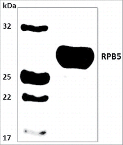
Figure 3. Western blot analysis on PVDF membrane. Lane 1, Molecular weight marker. Lane 2, Rpb5 recombinant protein detected by western blotting with a mouse anti-His antibody.

Figure 3. Western blot analysis on PVDF membrane. Lane 1, Molecular weight marker. Lane 2, Rpb5 recombinant protein detected by western blotting with a mouse anti-His antibody.

Reprints and Corporate Permissions

Please note: Selecting permissions does not provide access to the full text of the article, please see our help page How do I view content?

To request a reprint or corporate permissions for this article, please click on the relevant link below:

Academic Permissions

Please note: Selecting permissions does not provide access to the full text of the article, please see our help page How do I view content?

Obtain permissions instantly via Rightslink by clicking on the button below:

If you are unable to obtain permissions via Rightslink, please complete and submit this Permissions form. For more information, please visit our Permissions help page.