Figures & data
Figure 1. Coordinate system of hip joint model.
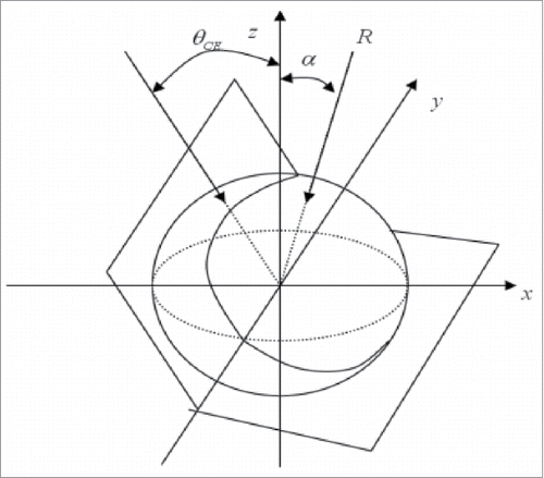
Figure 2. CE angle measurement.
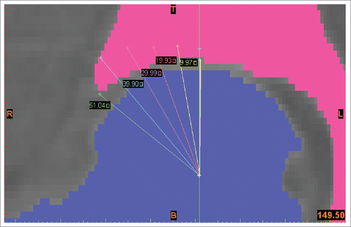
Figure 3. Finite element modeling process.
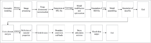
Figure 4. Finite element model of the hip joint.
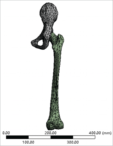
Table 1. Peak stress of different CE angle femoral head surface under different load direction.
Figure 5. Contrast of simulation results (actual value) and calculated results (theoretical value) in the mathematical model.
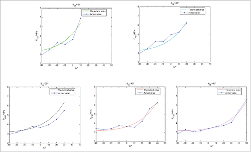
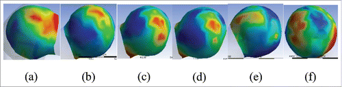
Figure 6. von Mises stress diagram in the articular surface of the hip joint. (A) von Mises stress diagram in the articular surface of the hip joint with the θCE = 50° and = 0°; (B) von Mises stress diagram in the articular surface of the hip joint with the θCE = 40° and
= −30°; (C) von Mises stress diagram in the articular surface of the hip joint with the θCE = 30° and
= −40°; (D) von Mises stress diagram in the articular surface of the hip joint with the θCE =30° and
= −50°; (E) von Mises stress diagram in the articular surface of the hip joint with the θCE = 20° and
=10°; (F) von Mises stress diagram in the articular surface of the hip joint with the θCE = 10° and
= −60°.