
Figures & data
Figure 1. (A) The umbrella-shaped sinus lift curette YSL-04 (MCT company). Inserted images showed the tips; (B) The probe-improved sinus lift curettes (designed and fabricated by the corresponding author of this paper). Inserted images showed the tips (b1 and b2 were used for detaching sinus mucosa in mesiodistal directions; b3 and b4 were used for detaching sinus mucosa in buccal-palatal directions); (C) The shape-memory Ni/Ti alloy wire containing tube elevator (elevator 014, invented and fabricated by the corresponding author of this paper) showing the primary detaching knife (the part between the 2 red lines in the picture).
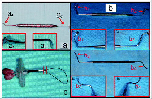
Figure 2. (A) The sketch of elevator 014 showing the main components (1: primary detaching knife; 2: tube; 3: holder; 4: spring wire; 5: scale; 6: scales; 7: stopper; 8: shape-memory Ni/Ti alloy wire.); (B) The sketch of primary detaching (10: implant hole or transcrestal access; 11: maxillary sinus mucosa.); and (C) The sketch of secondary detaching (9: residual alveolar bone).
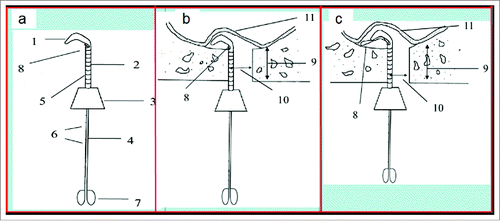
Figure 3. Goat ex vivo primary model showing the exposed maxillary sinus.
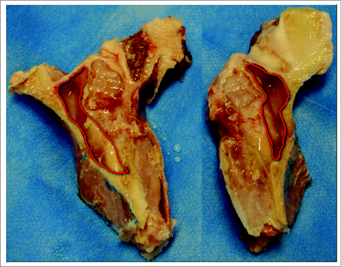
Figure 4. The determination of space volume created under the elevated maxillary sinus mucosa: (A) adding saline into the space before detaching maxillary sinus mucosa through the implant socket; (B) detaching maxillary sinus mucosa ex vivo using umbrella-shaped sinus lift curette YSL-04 with the tip of YSL-04 being seen under the sinus mucosa (red arrow); (C) detaching maxillary sinus mucosa ex vivo using probe-improved sinus lift curette with the tip being seen under the sinus mucosa (red arrow); (D) detaching maxillary sinus mucosa ex vivo using elevator 014 with the shape-memory Ni/Ti alloy wire being seen under the sinus mucosa (red arrow); (E) adding saline into the space after detaching maxillary sinus mucosa through the implant socket.
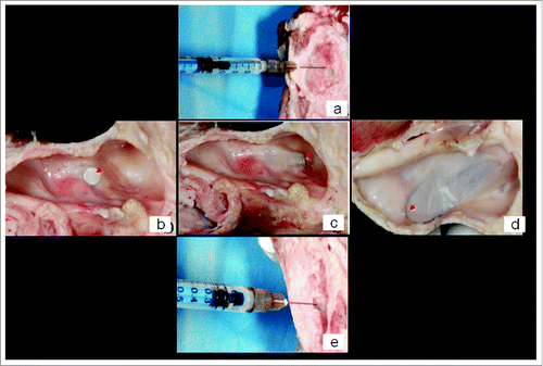
Table 1. The efficacy of ex vivo detaching goat maxillary sinus mucosa for 3 different sinus lift tools. Data were reported as mean ± standard deviation.