Figures & data
Figure 1. Models and ratios of tumor survival and apoptosis of the human PLC and HuH7 tumors in female nude mice. In order to estimate the differences between tumor survival and apoptosis, the polynomial area of the diagrams was calculated. Without viral therapy the tumors are in indefinite proliferation.
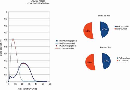
Figure 2. Models and ratios of tumor survival and apoptosis of the human PLC and HuH7 tumors in female nude mice after therapy with GLV-1h68 vaccinia virus strain. Again the polynomial areas of the diagrams were calculated. After virus therapy both tumor strains are moved into apoptosis.
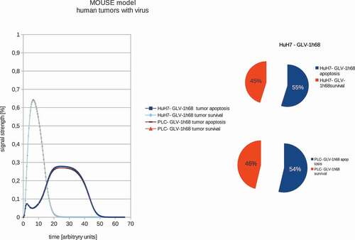
Figure 3. Models and ratios of tumor survival and apoptosis of the human PC-3 tumors in female nude mice without and with GLV-1h68-treatment. Without oncolytic virus therapy the tumor apoptosis rate (53%) is was set to be higher than the apoptotic rate (47%) and therefore simulating a proliferating tumor. After calculating the effects of viral therapy, these values ended up at 51% tumor survival and 49% tumor apoptosis and therefore ensuring tumor survival, albeit to a lesser extent.
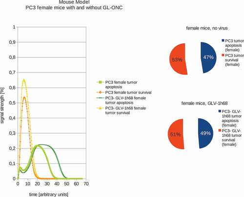
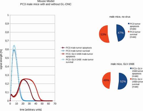
Figure 4. Models and ratios of tumor survival and apoptosis of the human PC-3 tumors in male nude mice without and with GLV-1h68-treatment. In stark contrast to the female mice, tumors in male mice react very differently to oncolytic virus therapy: after application of GLV-1h68 the tumor survival rate was calculated to be 48% with an apoptosis rate of 52% and therefore indicating tumor apoptosis.