Figures & data
Figure 1. Identification of A7r5 cells obtained from European collection of cell cultures.
(a) Primary cultures of A7r5 (phase contrast). (b) Co-staining of nuclei and α-smooth muscle actin (fluorescence/immunofluorescence)(200× magnification).
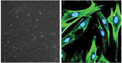
Figure 2. Effect of LOC285194 overexpression on proliferation and apoptosis in A7r5 cells in vitro.
(a) A7r5 cells were infected with Lv-LOC285194 or Lv-NC for 72 h. (a) LOC285194 expression was detacted by qPCR assay; (b) A7r5 cells were co-infected with Lv-LOC285194/Lv-NC and PUMA siRNA, cell apoptosis was detected by ELISA. (c) cell apoptosis was detected by TUNEL staining. (d) cell proliferation was detected by CCK-8 assay; *p < 0.05; **p < 0.01; ***p < 0.001.
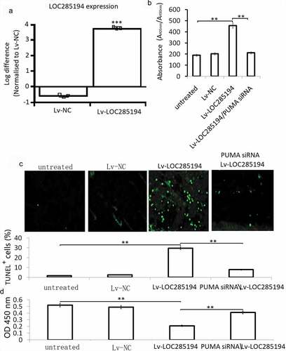
Figure 3. Effect of LOC285194 overexpression on apoptotic and metastatic protein expression in vitro.
(a) A7r5 cells were infected with Lv-LOC285194 or Lv-NC for 72 h, pro- and anti-apoptotic protein expression were detected by Western blot assay. (b) A7r5 cells were infected with Lv-LOC285194 or Lv-NC for 72 h, S100A4, MMP-2/9 were detected by western blot assay; A7r5 cells were infected with S100A4 siRNA or NC siRNA for 24 h, S100A4 protein expression was detected by western blot assay.
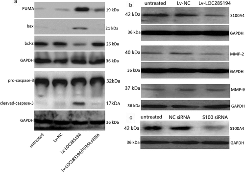
Figure 4. Effect of LOC285194 overexpression and S100A4 downexpression on invasion and migration in vitro.
(a,b) A7r5 cells were infected with Lv-LOC285194 or S100A4 siRNA and its control for 24 h; cell invasion and migration were detected using Transwell assay; (c,d) A7r5 cells were infected with Lv-LOC285194 or S100A4 siRNA and its control for 24 h and 48 h; cell migration was detected using Wound healing assay.*p < 0.05.
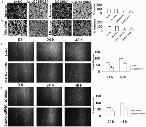
Figure 5. Effect of miR-211 on LOC285194 -induced PUMA expression.
(a) The A7r5 cells were transfected with Lv-LOC285194 or co-transfected with miR-211 mimic or its control miR-NC for 72 h. miR-211 expression was detected by qPCR assay; (b) The A7r5 cells were transfected with miR-211 inhibitor or its control NC inhibitor for 72 h. miR-211 expression was detected by qPCR assay; (c) PUMA protein expression was detected by western blot assay. (d) The A7r5 cells were transfected with Lv-LOC285194 or co-transfected with miR-211 mimic or its control miR-NC for 72 h. PUMA protein expression was detected by western blot assay.
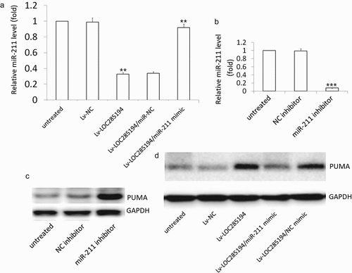
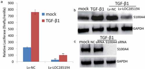
Figure 6. TGF-β1 is required for S100A4 inhibition by LOC285194.
(a) A7r5 cells were Lv-LOC285194 or Lv-NC (control) and then treated with TGF-β1 (5 ng/ml; 24 h) (+) or mock treated (-), Luciferase is expressed as a ratio of firefly/renilla. (b) S100A4 was assessed by Western blot. (c) A7r5 cells were transfected with S100A4 siRNA or control siRNA for 48 h, then treated with TGF-β1 (5 ng/ml; 24 h), S100A4 was assessed by Western blot.**p < 0.01.