
Figures & data
Figure 1. ATF3 expression is up-regulated in human keloid tissues
The expression levels of ATF3 in the healthy controls, keloid tissue and the normal skin were determined by real-time PCR (a), western blot (b) and immunohistochemistry (c, 400X). *** P < 0.001.
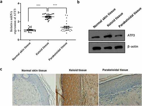
Figure 2. ATF3 promotes cell proliferation and collagen production in keloid fibroblast cells
(a and b) Western blot confirmed that ATF3 was upregulated or downregulated in keloid fibroblast cells following transfection with recombinant vectors encoding ATF3 or siRNA targeting ATF3. (c) The cell viability of fibroblast cells was evaluated by MTT assay in different groups. Real-time PCR was performed to determine the mRNA expression of TGF-β1 (d), FGF2 (e), FGF8 (f), COLⅠ (g) and COLⅠ (h). (i) Western blot was conducted to examined the protein levels of TGF-β1, FGF2, FGF8, COLⅠ and COLⅠ in different groups. ** P < 0.01; *** P < 0.001, compared to control.
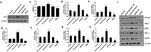
Figure 3. ATF3 suppresses apoptosis in keloid fibroblast cells
Keloid fibroblast cells were transfected with recombinant vectors encoding ATF3 or siRNA targeting ATF3. Flow cytometry was used to detect the cell apoptosis. In addition, real-time PCR (b) and western blot (c) were conducted to detect the mRNA and protein expression of BCL2, Bad, Caspase3 and Caspase9. *** P < 0.001, compared to control.

Figure 4. ATF3 inhibited the expression of pro-apoptosis factors in keloid fibroblast cells
Keloid fibroblast cells were transfected with recombinant vectors encoding ATF3 or siRNA targeting ATF3. Real-time PCR was conducted to detect the mRNA expression of BCL2 (a), Bad (b), Caspase3 (c) and Caspase9 (d) in fibroblast cells. Western blot (e) was performed to determine the protein expression of BCL2, Bad, Caspase3 and Caspase9 in fibroblast cells. *** P < 0.001, compared to control.
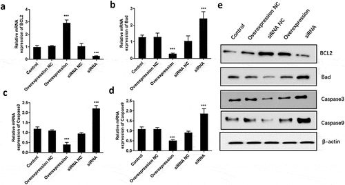
Figure 5. ATF3 promotes the invasive potential of keloid fibroblast cells
Keloid fibroblast cells were transfected with recombinant vectors encoding ATF3 or siRNA targeting ATF3. Transwell invasion assay was used to evaluate the invasive potential of keloid fibroblast cells. Moreover, real-time PCR (b) and western blot (c) were conducted to detect the mRNA and protein expression of MMP1, MMP2, MMP9 and MMP13. *** P < 0.001, compared to control.
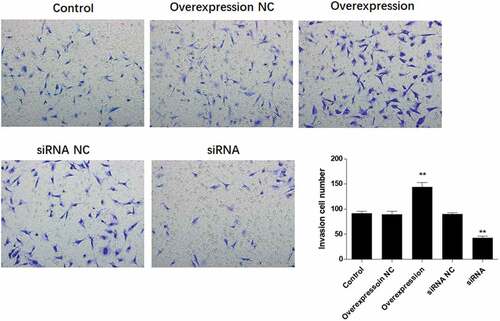
Figure 6. ATF3 promotes the expression of MMPs in keloid fibroblast cells
Keloid fibroblast cells were transfected with recombinant vectors encoding ATF3 or siRNA targeting ATF3. Real-time PCR was performed to determine the mRNA expression of MMP1 (a), MMP2 (b), MMP9 (c) and MMP13 (d) in fibroblast cells. Western blot (e) was conducted to detect the protein expression MMP1/2/8/13 in fibroblast cells. *** P < 0.001, compared to control.
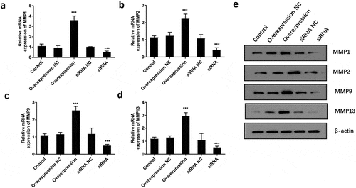
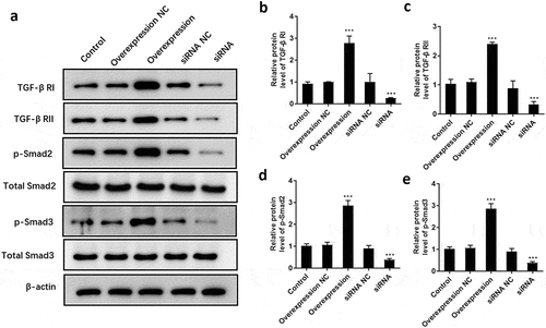
Figure 7. ATF3 promotes the activation of TGF-β/Smad signaling pathway in keloid fibroblast cells
Keloid fibroblast cells were transfected with recombinant vectors encoding ATF3 or siRNA targeting ATF3. (a) Western blot was conducted to detect the protein levels of TGF-β RI, TGF-β RII, p-Smad2, p-Smad3, total Smad2, and total Smad3. Quantification of protein signals of TGF-β RI (b) TGF-β RII (c), p-Smad2 (d), and p-Smad3 (e). *** P < 0.001, compared to control.
Supplemental material