Figures & data
Table 1. Synthetic siRNA sequences
Table 2. qPCR primer sequences
Figure 1. The expression of PGM5P4-AS1 was increased in lung cancer tissues. (a), Boxplot of PGM5P4-AS1 in LUAD and LUSC; LUAD, lung adenocarcinoma; LUSC, lung squamous cell carcinoma; FC, fold change (*p < 0.05 vs. normal tissues). Blue: tumor tissue; black: normal tissue. (b-c), Relative expression levels of PGM5P4-AS1 were measured by qRT-PCR assay in lung cancer tissues and adjacent tissues, as well as lung cancer cell lines, respectively (&p < 0.05 vs. adjacent tissues). (d-e), Transfection efficiency was detected after A549 and SK-MES-1 cells were transfected with PGM5P4-AS1 siRNA or PGM5P4-AS1-OE for 48 h, respectively (**p < 0.01 vs. NC siRNA, ##p < 0.01 vs. vector)
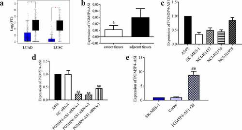
Figure 2. PGM5P4-AS1 suppressed the proliferation of lung cancer cells. (a-b), Cell proliferation of A549 and SK-MES-1 cells was measured by MTT assay after being transfected with PGM5P4-AS1 siRNA or PGM5P4-AS1-OE for 0 h, 24 h, 48 h, 72 h, or 96 h (#p < 0.05 vs. vector, ##p < 0.01 vs. vector, **p<0.01 vs. NC siRNA). (c-d), Cell cycles arrest of A549 and SK-MES-1 cells was measured by flow cytometer after being transfected with PGM5P4-AS1 siRNA or PGM5P4-AS1-OE for 48 h (#p < 0.05 vs. vector, ##p<0.01 vs. vector, **p<0.01 vs. NC siRNA)
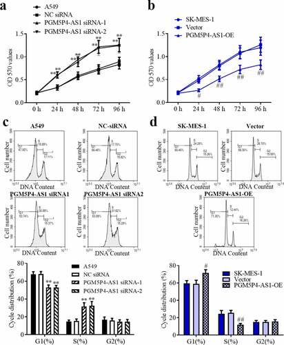
Figure 3. PGM5P4-AS1 inhibited the in vivo growth of lung cancer. (a), PGM5P4-AS1-OE or untreated vectors were injected in nude mice with xenograft tumors. (b), Cell proliferation ability of tumor was evaluated via ki67 immunohistochemistry (×400). The scale bar represented 50 μm. (c), Volume of the excised tumor (#p < 0.05 vs. vector, ##p < 0.01 vs. vector). (d), Weight of the excised tumor (##p < 0.01 vs. vector). (e), Relative expression of PGM5P4-AS1 by qRT-PCR in the tumor (##p < 0.01 vs. vector)
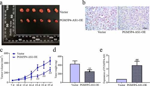
Figure 4. PGM5P4-AS1 inhibited the migration and invasion activities of lung cancer cells. (a), The migration ability of A549 cells was assessed by wound healing assay, and the results showed that interference in the expression of PGM5P4-AS1 could promote cell migration (×100, *p < 0.05 vs. NC siRNA). The scale bar represented 200 μm. (b), The results of the migration ability of SK-MES-1 cells showed that the overexpression of PGM5P4-AS1 could inhibit cell migration (×100, #p < 0.05 vs. vector). The scale bar represented 200 μm. (c), Invasion abilities were measured by transwell assay, the results showed that interference in the expression of PGM5P4-AS1 could promote cell invasion (×200, *p < 0.05 vs. NC siRNA). The scale bar represented 100 μm. (d), The overexpression of PGM5P4-AS1 could inhibit the cell invasion (×200, #p < 0.05 vs. vector). The scale bar represented 100 μm
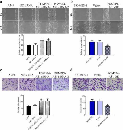
Figure 5. PGM5P4-AS1 decreased the miR-1275 level and promoted the expression of LZTS3. (a-b), Relative expression levels of miRNA-1275 and LZTS3 in A549 cells, that were interfered in PGM5P4-AS1 expression (**p < 0.01 vs. NC siRNA). (c-d), Relative expression levels of miRNA-1275 and LZTS3 in SK-MES-1 cells, that were overexpressed of PGM5P4-AS1 (##p < 0.01 vs. vector). (e-f), Protein expression levels of LZTS3, cyclinD1, MMP-2, Bcl-2, and Bcl-xl in A549 cells, that were transfected with PGM5P4-AS1 siRNA or PGM5P4-AS1-OE, respectively (**p < 0.01 vs. NC siRNA, ##p < 0.01 vs. vector)
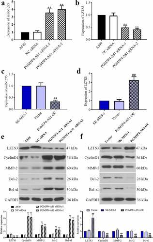
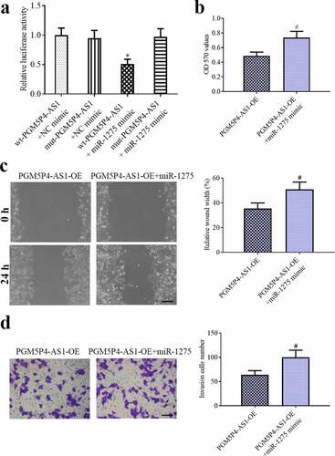
Figure 6. PGM5P4-AS1 suppressed the malignant behavior of lung cancer cells by sponging miR-1275. (a), The Dual-luciferase reporter gene showed that PGM5P4-AS1 sponged miR-1275 (*p < 0.05 vs. wt-PGM5P4-AS1+ NC mimic). (b), MTT assay was performed to evaluate the ability of cell proliferation (#p < 0.05 vs. PGM5P4-AS1-OE). (c), the migration ability of cells was assessed by wound healing assay (×100, Scale bar represented 200 μm; #p < 0.05 vs. PGM5P4-AS1-OE). (d), Transwell assay was used to estimate the cell invasion ability (×200, Scale bar represented 100 μm; #p < 0.05 vs. PGM5P4-AS1-OE)